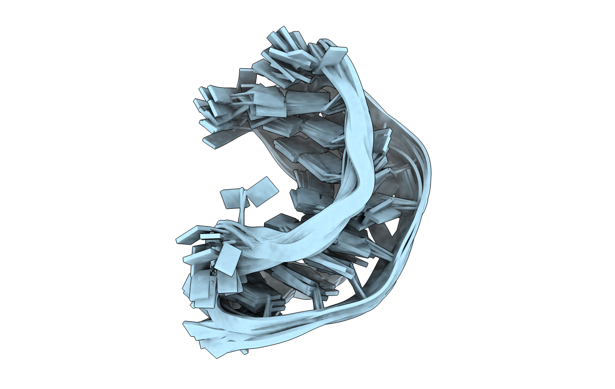
1KPY image
Deposition Date 2002-01-03
Release Date 2002-01-11
Last Version Date 2024-05-01
Entry Detail
PDB ID:
1KPY
Keywords:
Title:
PEMV-1 P1-P2 Frameshifting Pseudoknot, 15 Lowest Energy Structures
Biological Source:
Source Organism:
Method Details:
Experimental Method:
Conformers Calculated:
100
Conformers Submitted:
15
Selection Criteria:
structures with the lowest energy
Macromolecular Entities
Polymer Type:polyribonucleotide
Molecule:P1-P2 frameshifting pseudoknot
Chain IDs:A
Chain Length:33
Number of Molecules:1
Biological Source:
Modified Residue
Compound ID Chain ID Parent Comp ID Details 2D Image
CH A C N3-PROTONATED CYTIDINE-5'-MONOPHOSPHATE
Ligand Molecules
Primary Citation
Solution structure of a luteoviral P1-P2 frameshifting mRNA pseudoknot
J.Mol.Biol. 322 621 633 (2002)
PMID: 12225754 DOI: 10.1016/S0022-2836(02)00779-9

Abstact

A hairpin-type messenger RNA pseudoknot from pea enation mosaic virus RNA1 (PEMV-1) regulates the efficiency of programmed -1 ribosomal frameshifting. The solution structure and 15N relaxation rates reveal that the PEMV-1 pseudoknot is a compact-folded structure composed almost entirely of RNA triple helix. A three nucleotide reverse turn in loop 1 positions a protonated cytidine, C(10), in the correct orientation to form an A((n-1)).C(+).G-C(n) major groove base quadruple, like that found in the beet western yellows virus pseudoknot and the hepatitis delta virus ribozyme, despite distinct structural contexts. A novel loop 2-loop 1 A.U Hoogsteen base-pair stacks on the C(10)(+).G(28) base-pair of the A(12).C(10)(+).G(28)-C(13) quadruple and forms a wedge between the pseudoknot stems stabilizing a bent and over-rotated global conformation. Substitution of key nucleotides that stabilize the unique conformation of the PEMV-1 pseudoknot greatly reduces ribosomal frameshifting efficacy.

Legend

Protein

Chemical

Disease

Primary Citation of related structures