1KP0 image
Deposition Date 2001-12-26
Release Date 2002-07-31
Last Version Date 2024-10-09
Entry Detail
PDB ID:
1KP0
Keywords:
Title:
The Crystal Structure Analysis of Creatine Amidinohydrolase from Actinobacillus
Biological Source:
Source Organism:
Actinobacillus (Taxon ID: 713)
Method Details:
Experimental Method:
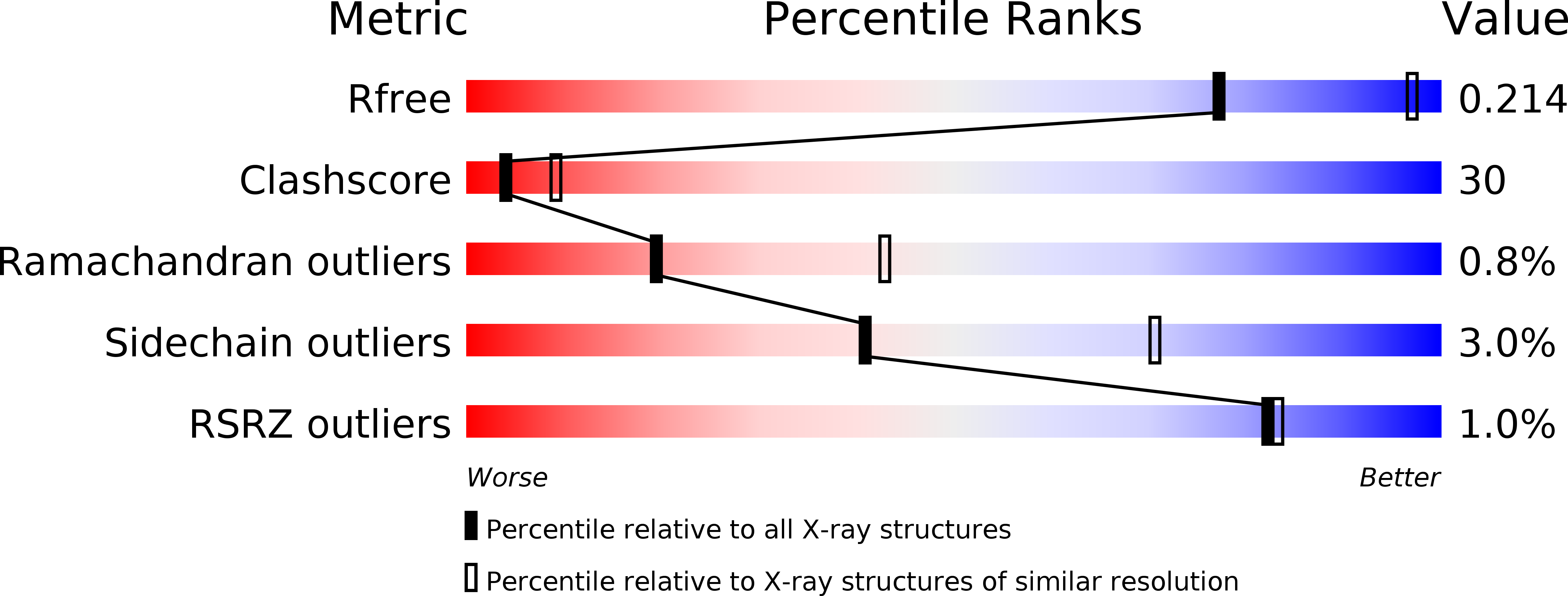
Resolution:
2.70 Å
R-Value Free:
0.22
R-Value Work:
0.18
R-Value Observed:
0.19
Space Group:
I 2 2 2
Macromolecular Entities
Structures with similar UniProt ID
Protein Blast
Polymer Type:polypeptide(L)
Molecule:CREATINE AMIDINOHYDROLASE
Chain IDs:A, B
Chain Length:402
Number of Molecules:2
Biological Source:Actinobacillus
Primary Citation
Structure of creatine amidinohydrolase from Actinobacillus.
Acta Crystallogr.,Sect.D 58 1322 1328 (2002)
PMID: 12136144 DOI: 10.1107/S0907444902010156

Abstact

The crystal structure of Actinobacillus creatine amidinohydrolase has been solved by molecular replacement. The amino-acid sequence has been derived from the crystal structure. Crystals belong to space group I222, with unit-cell parameters a = 111.26 (3), b = 113.62 (4), c = 191.65 (2) A, and contain two molecules in an asymmetric unit. The structure was refined to an R factor of 18.8% at 2.7 A resolution. The crystal structure contains a dimer of 402 residues and 118 water molecules. The protein structure is bilobal, consisting of a small N-terminal domain and a large C-terminal domain. The C-terminal domain has a pitta-bread fold, similar to that found in Pseudomonas putida creatinase, proline aminopeptidases and methionine aminopeptidase. Comparison with complex crystal structures of P. putida creatinase reveals that the enzyme activity of Actinobacillus creatinase might be similar to that of P. putida creatinase.

Legend

Protein

Chemical

Disease

Primary Citation of related structures