1KN3 image
Deposition Date 2001-12-18
Release Date 2002-06-12
Last Version Date 2023-08-16
Entry Detail
PDB ID:
1KN3
Keywords:
Title:
Murine PEBP-2 (phosphatidylethanolamine-binding protein-2)
Biological Source:
Source Organism:
Mus musculus (Taxon ID: 10090)
Method Details:
Experimental Method:
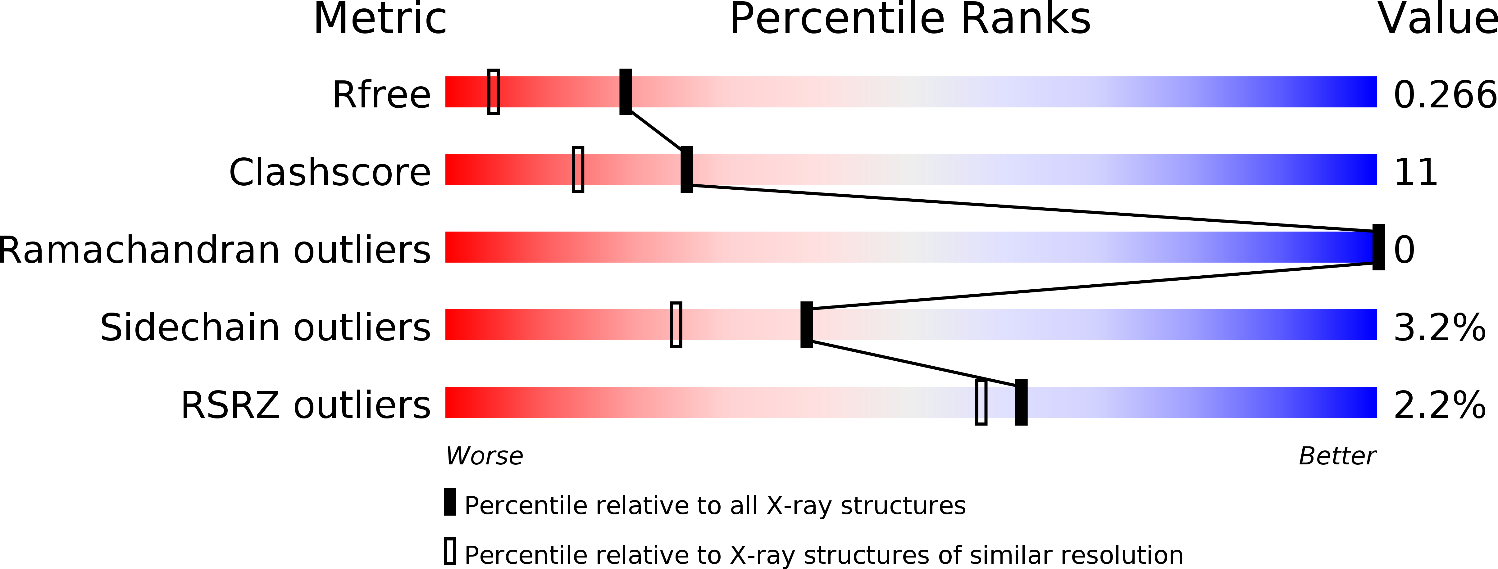
Resolution:
1.80 Å
R-Value Free:
0.26
R-Value Work:
0.21
R-Value Observed:
0.21
Space Group:
P 21 21 21
Macromolecular Entities
Structures with similar UniProt ID
Protein Blast
Polymer Type:polypeptide(L)
Molecule:Phosphatidylethanolamine Binding Protein-2
Gene (Uniprot):Pbp2
Chain IDs:A
Chain Length:183
Number of Molecules:1
Biological Source:Mus musculus
Primary Citation
The crystal structure of PEBP-2, a homologue of the PEBP/RKIP family.
Acta Crystallogr.,Sect.D 58 1077 1080 (2002)
PMID: 12037323 DOI: 10.1107/S090744490200522X

Abstact

Proteins from the PEBP (phosphatidylethanolamine-binding protein) family have been identified in a wide variety of species and are thought to regulate a range of intracellular signalling cascades. The rat homologue (known as RKIP; Raf-1 kinase inhibitor protein) has been shown to negatively regulate the MAP kinase pathway through formation of inhibitory complexes with Raf-1 and MEK. The crystal structure of a new, murine member of the PEBP family, termed mPEBP-2, has been determined. On the basis of amino-acid homology, mPEBP-2 belongs to a distinct subset of the mammalian PEBP proteins. Nonetheless, mPEBP-2 is seen to be very similar in structure to other PEBP proteins from human, bovine and plant sources. Regions of distinctive sequence associated with the PEBP-2 subset are discussed with reference to this structure.

Legend

Protein

Chemical

Disease

Primary Citation of related structures