
Deposition Date
2001-12-17
Release Date
2002-02-20
Last Version Date
2023-08-16
Entry Detail
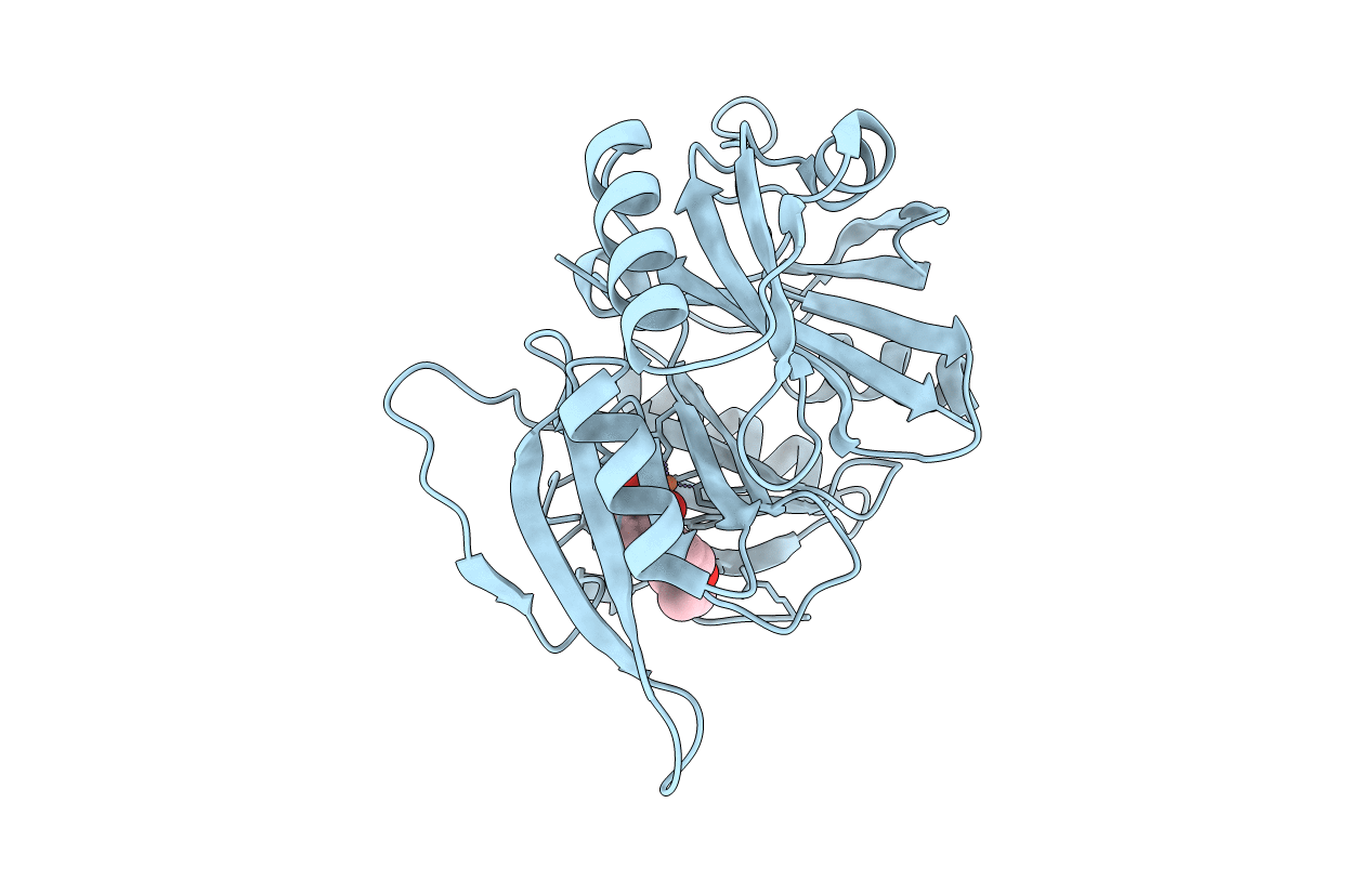
PDB ID:
1KMY
Keywords:
Title:
Crystal Structure of 2,3-dihydroxybiphenyl 1,2-dioxygenase Complexed with 2,3-dihydroxybiphenyl under Anaerobic Condition
Biological Source:
Source Organism(s):
Burkholderia xenovorans (Taxon ID: 266265)
Expression System(s):
Method Details:
Experimental Method:
Resolution:
2.00 Å
R-Value Free:
0.19
R-Value Work:
0.16
R-Value Observed:
0.19
Space Group:
I 4 2 2
