
Deposition Date
2001-12-17
Release Date
2002-07-24
Last Version Date
2024-10-09
Entry Detail
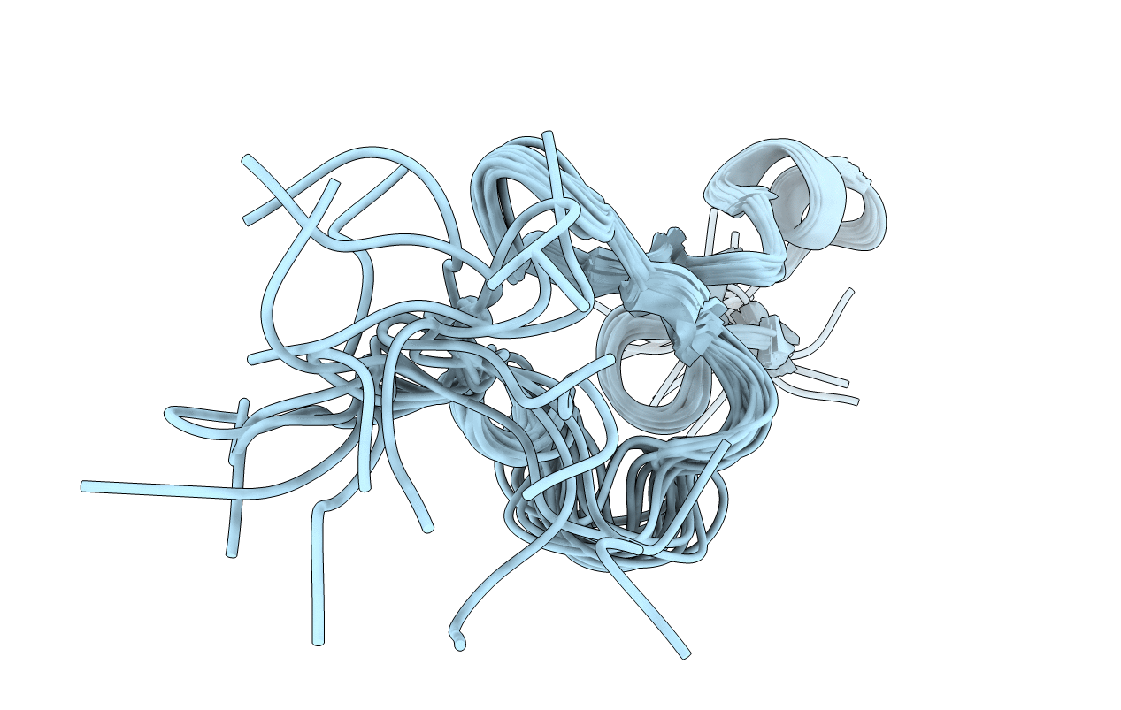
PDB ID:
1KMX
Keywords:
Title:
Heparin-binding Domain from Vascular Endothelial Growth Factor
Biological Source:
Source Organism(s):
Homo sapiens (Taxon ID: 9606)
Expression System(s):
Method Details:
Experimental Method:
Conformers Calculated:
100
Conformers Submitted:
20
Selection Criteria:
structures with the least restraint violations
