
Deposition Date
2001-12-17
Release Date
2002-07-10
Last Version Date
2024-04-03
Entry Detail
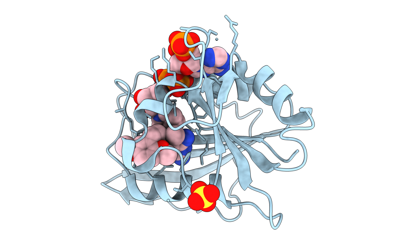
PDB ID:
1KMV
Keywords:
Title:
HUMAN DIHYDROFOLATE REDUCTASE COMPLEXED WITH NADPH AND (Z)-6-(2-[2,5-DIMETHOXYPHENYL]ETHEN-1-YL)-2,4-DIAMINO-5-METHYLPYRIDO[2,3-D]PYRIMIDINE (SRI-9662), A LIPOPHILIC ANTIFOLATE
Biological Source:
Source Organism(s):
Homo sapiens (Taxon ID: 9606)
Expression System(s):
Method Details:
Experimental Method:
Resolution:
1.05 Å
R-Value Free:
0.18
R-Value Work:
0.13
Space Group:
P 21 21 21
