Deposition Date
2001-12-14
Release Date
2002-02-13
Last Version Date
2024-11-20
Entry Detail
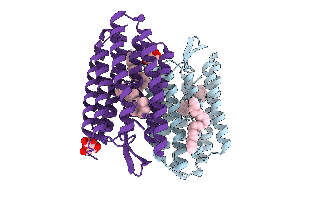
PDB ID:
1KME
Keywords:
Title:
CRYSTAL STRUCTURE OF BACTERIORHODOPSIN CRYSTALLIZED FROM BICELLES
Biological Source:
Source Organism(s):
Halobacterium salinarum (Taxon ID: 2242)
Method Details:
Experimental Method:
Resolution:
2.00 Å
R-Value Free:
0.27
R-Value Work:
0.26
R-Value Observed:
0.26
Space Group:
P 1 21 1