Deposition Date
2001-12-11
Release Date
2002-06-05
Last Version Date
2024-10-30
Entry Detail
PDB ID:
1KLF
Keywords:
Title:
FIMH ADHESIN-FIMC CHAPERONE COMPLEX WITH D-MANNOSE
Biological Source:
Source Organism(s):
Escherichia coli (Taxon ID: 562)
Expression System(s):
Method Details:
Experimental Method:
Resolution:
2.79 Å
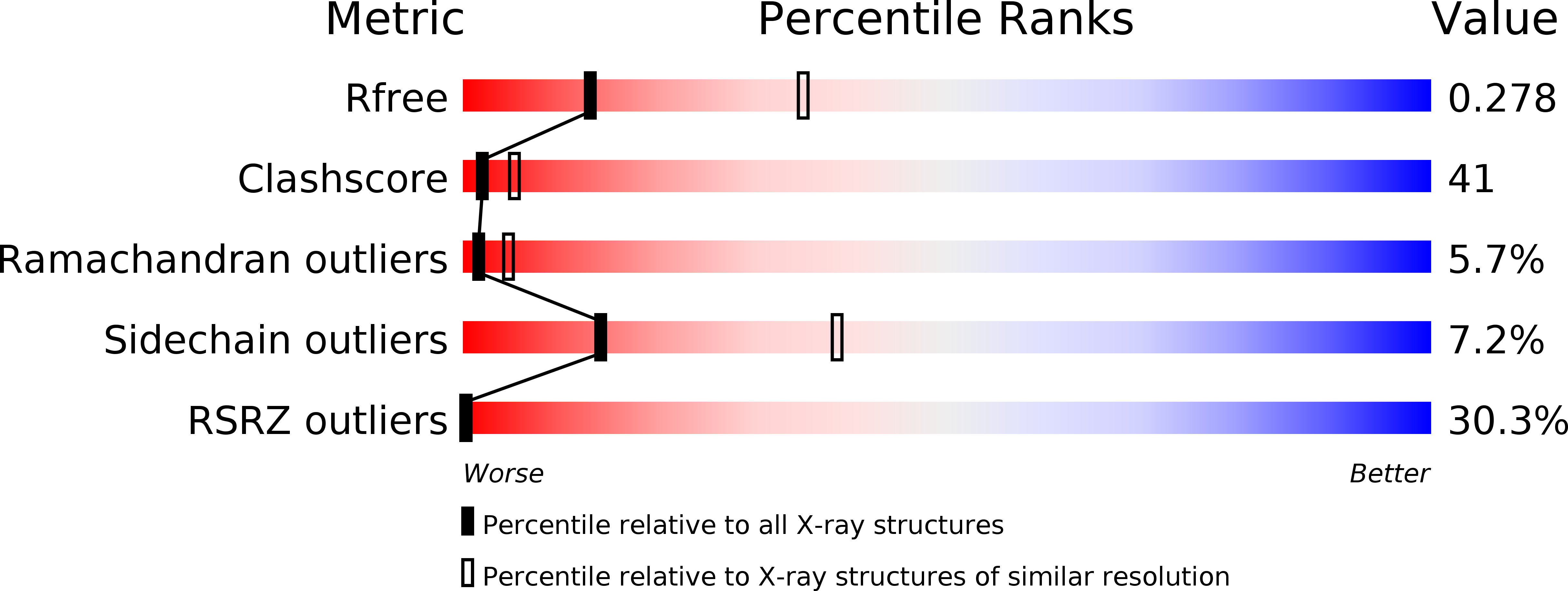
R-Value Free:
0.28
R-Value Work:
0.23
R-Value Observed:
0.23
Space Group:
C 1 2 1