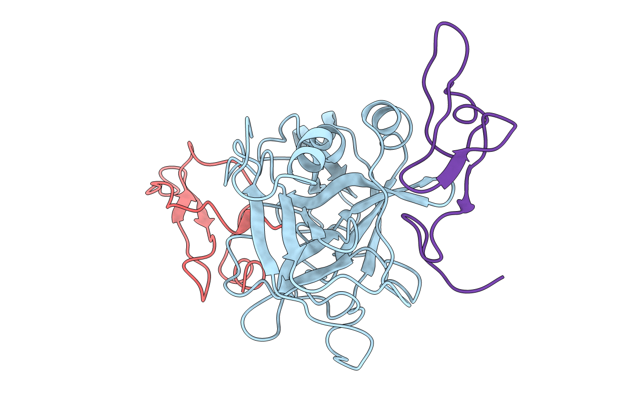
1KIG image
Deposition Date 1997-04-24
Release Date 1998-10-28
Last Version Date 2024-10-23
Entry Detail
PDB ID:
1KIG
Title:
BOVINE FACTOR XA
Biological Source:
Source Organism:
Bos taurus (Taxon ID: 9913)
Ornithodoros moubata (Taxon ID: 6938)
Method Details:
Experimental Method:
Resolution:
3.00 Å
R-Value Work:
0.18
R-Value Observed:
0.18
Space Group:
P 42 21 2
Macromolecular Entities
Structures with similar UniProt ID
Protein Blast
Polymer Type:polypeptide(L)
Molecule:FACTOR XA
Gene (Uniprot):F10
Chain IDs:A (auth: H)
Chain Length:241
Number of Molecules:1
Biological Source:Bos taurus
Structures with similar UniProt ID
Protein Blast
Polymer Type:polypeptide(L)
Molecule:ANTICOAGULANT PEPTIDE
Chain IDs:C (auth: I)
Chain Length:60
Number of Molecules:1
Biological Source:Ornithodoros moubata
Structures with similar UniProt ID
Protein Blast
Polymer Type:polypeptide(L)
Molecule:FACTOR XA
Gene (Uniprot):F10
Chain IDs:B (auth: L)
Chain Length:51
Number of Molecules:1
Biological Source:Bos taurus
Ligand Molecules
Primary Citation
Unexpected binding mode of tick anticoagulant peptide complexed to bovine factor Xa.
J.Mol.Biol. 283 147 154 (1998)
PMID: 9761680 DOI: 10.1006/jmbi.1998.2069

Abstact

The structure of recombinant tick anticoagulant peptide (rTAP) complexed to bovine factor Xa at 3.0 A resolution reveals the structural basis for the specificity and the high affinity of rTAP. Three N-terminal residues, Tyr501, Asn502 and Arg503, play a critical role in the complex formation as suggested by earlier mutagenic studies and the ornithodorin-thrombin complex. Unexpectedly, the side-chain of Tyr501 is located in the S1 pocket, although factor Xa favors arginine as a P1 residue. Arg503 is located at the aryl binding pocket and forms a salt-bridge with Glu97 of factor Xa. The autolysis loop, which is disordered in the uninhibited factor Xa structure, is involved in the formation of the complex as a part of the secondary binding site. The C-terminal helix of rTAP interacts with factor Xa as a secondary binding determinant. The N-terminal residues of rTAP reorganize during the formation of the factor Xa-rTAP complex from the conformation found in the solution into an extended conformation. The presence of the secondary binding site confirms the proposed two-step kinetic mechanism based on the results of a mutagenesis study.

Legend

Protein

Chemical

Disease

Primary Citation of related structures