
Deposition Date
2001-12-03
Release Date
2002-05-15
Last Version Date
2023-08-16
Entry Detail
PDB ID:
1KIE
Keywords:
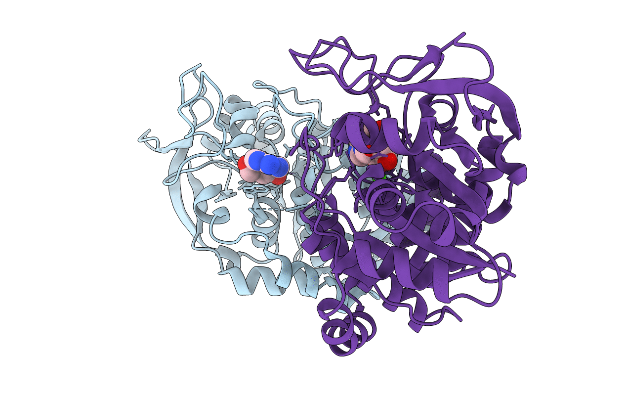
Title:
Inosine-adenosine-guanosine preferring nucleoside hydrolase from Trypanosoma vivax: Asp10Ala mutant in complex with 3-deaza-adenosine
Biological Source:
Source Organism(s):
Trypanosoma vivax (Taxon ID: 5699)
Expression System(s):
Method Details:
Experimental Method:
Resolution:
2.00 Å
R-Value Free:
0.21
R-Value Work:
0.15
Space Group:
P 1 21 1
