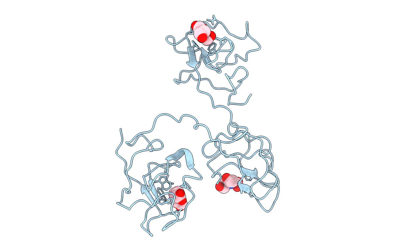
1KI0 image
Deposition Date 2001-12-02
Release Date 2002-05-29
Last Version Date 2024-11-06
Entry Detail
PDB ID:
1KI0
Keywords:
Title:
The X-ray Structure of Human Angiostatin
Biological Source:
Source Organism:
Homo sapiens (Taxon ID: 9606)
Host Organism:
Method Details:
Experimental Method:
Resolution:
1.75 Å
R-Value Free:
0.26
R-Value Work:
0.19
Space Group:
P 41 21 2
Macromolecular Entities
Polymer Type:polypeptide(L)
Molecule:ANGIOSTATIN
Gene (Uniprot):PLG
Mutations:N289E
Chain IDs:A
Chain Length:253
Number of Molecules:1
Biological Source:Homo sapiens
Ligand Molecules
Primary Citation
The X-ray crystallographic structure of the angiogenesis inhibitor angiostatin.
J.Mol.Biol. 318 1009 1017 (2002)
PMID: 12054798 DOI: 10.1016/S0022-2836(02)00211-5

Abstact

Angiogenesis inhibitors have gained much public attention recently as anti-cancer agents and several are currently in clinical trials, including angiostatin (Phase I, Thomas Jefferson University Hospital, Philadelphia, PA). We report here the bowl-shaped structure of angiostatin kringles 1-3, the first multi-kringle structure to be determined. All three kringle lysine-binding sites contain a bound bicine molecule of crystallization while the former of kringle 2 and kringle 3 are cofacial. Moreover, the separation of the kringle 2 and kringle 3 lysiner binding sites is sufficient to accommodate the alpha-helix of the 30 residue peptide VEK-30 found in the kringle 2/VEK-30 complex. Together the three kringles produce a central cavity suggestive of a unique domain where they may function in concert.

Legend

Protein

Chemical

Disease

Primary Citation of related structures