Deposition Date
2001-11-28
Release Date
2002-10-23
Last Version Date
2024-02-14
Entry Detail
PDB ID:
1KGZ
Keywords:
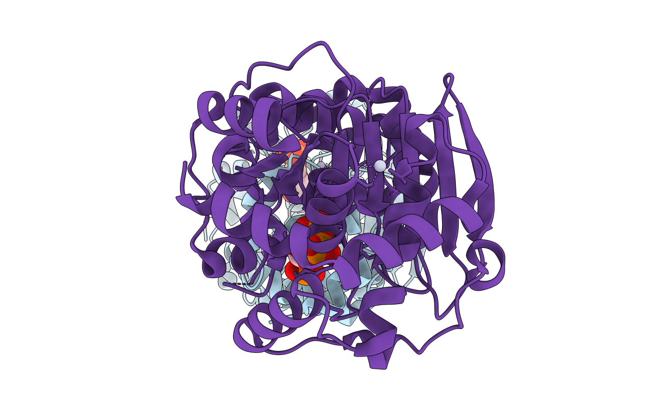
Title:
Crystal Structure Analysis of the Anthranilate Phosphoribosyltransferase from Erwinia carotovora (current name, Pectobacterium carotovorum)
Biological Source:
Source Organism(s):
Pectobacterium carotovorum (Taxon ID: 554)
Expression System(s):
Method Details:
Experimental Method:
Resolution:
2.40 Å
R-Value Free:
0.26
R-Value Work:
0.21
Space Group:
P 21 21 2