
Deposition Date
2001-11-16
Release Date
2001-12-21
Last Version Date
2024-10-16
Entry Detail
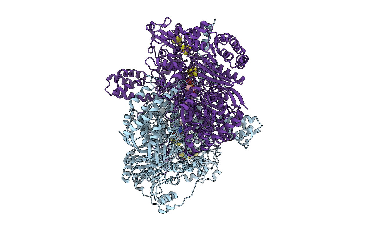
PDB ID:
1KEK
Keywords:
Title:
Crystal Structure of the Free Radical Intermediate of Pyruvate:Ferredoxin Oxidoreductase
Biological Source:
Source Organism(s):
Desulfovibrio africanus (Taxon ID: 873)
Method Details:
Experimental Method:
Resolution:
1.90 Å
R-Value Free:
0.22
R-Value Work:
0.17
R-Value Observed:
0.17
Space Group:
P 21 21 21
