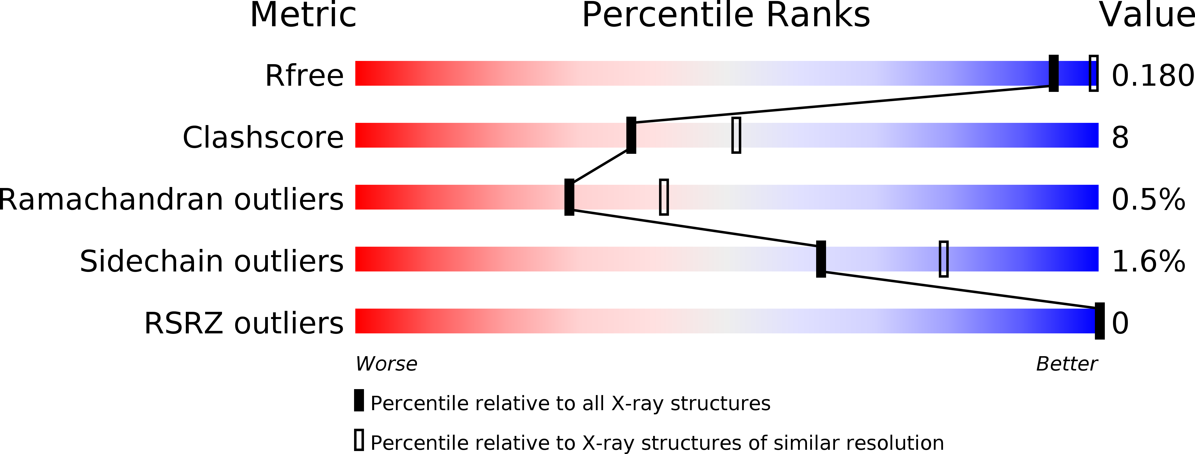
1KEG image
Deposition Date 2001-11-15
Release Date 2002-11-15
Last Version Date 2024-11-20
Entry Detail
PDB ID:
1KEG
Title:
Antibody 64M-2 Fab complexed with dTT(6-4)TT
Biological Source:
Source Organism:
Mus musculus (Taxon ID: 10090)
Method Details:
Experimental Method:
Resolution:
2.40 Å
R-Value Free:
0.26
R-Value Work:
0.20
R-Value Observed:
0.20
Space Group:
P 21 21 21
Macromolecular Entities
Polymer Type:polydeoxyribonucleotide
Molecule:5'-D(*TP*(64T)P*TP*T)-3'
Chain IDs:A
Chain Length:4
Number of Molecules:1
Biological Source:
Structures with similar UniProt ID
Protein Blast
Polymer Type:polypeptide(L)
Molecule:Anti-(6-4) photoproduct antibody 64M-2 Fab (heavy chain)
Chain IDs:C (auth: H)
Chain Length:220
Number of Molecules:1
Biological Source:Mus musculus
Polymer Type:polypeptide(L)
Molecule:Anti-(6-4) photoproduct antibody 64M-2 Fab (light chain)
Chain IDs:B (auth: L)
Chain Length:216
Number of Molecules:1
Biological Source:Mus musculus
Modified Residue
Compound ID Chain ID Parent Comp ID Details 2D Image
64T A DT 5-HYDROXY-THYMIDINE-5'-MONOPHOSPHATE
Ligand Molecules
Primary Citation
Structure of the DNA (6-4) photoproduct dTT(6-4)TT in complex with the 64M-2 antibody Fab fragment implies increased antibody-binding affinity by the flanking nucleotides.
Acta Crystallogr.,Sect.D 68 232 238 (2012)
PMID: 22349224 DOI: 10.1107/S0907444912000327

Abstact

Pyrimidine (6-4) pyrimidone DNA photoproducts produced by ultraviolet light are highly mutagenic and carcinogenic. The crystal structure of the dTT(6-4)TT photoproduct in complex with the Fab fragment of the antibody 64M-2 that is specific for (6-4) photoproducts was determined at 2.4 Å resolution. The dT(6-4)T segment is fully accommodated in the concave binding pocket of the Fab, as observed in the complex of dT(6-4)T with the Fab. The pyrimidine and pyrimidone bases of the dT(6-4)T segment are positioned nearly perpendicularly to each other. The thymidine segments flanking both ends extend away from the dT(6-4)T segment. The 5'-side thymine base is parallel to the side chain of Tyr100iH of the antibody heavy chain and is also involved in electrostatic interactions with Asn30L, Tyr32L and Lys50L of the antibody light chain. The 5'-side and 3'-side phosphate groups exhibit electrostatic interactions with Asn28L and Ser58H, respectively. These interactions with the flanking nucleotides explain why longer oligonucleotides containing dT(6-4)T segments in the centre show higher antibody-binding affinities than the dT(6-4)T ligand.

Legend

Protein

Chemical

Disease

Primary Citation of related structures