Deposition Date
1994-08-25
Release Date
1995-02-27
Last Version Date
2024-02-07
Entry Detail
PDB ID:
1KDC
Keywords:
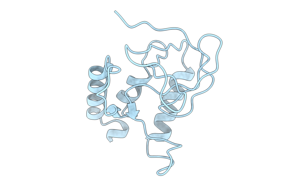
Title:
STABILIZATION OF A STRAINED PROTEIN LOOP CONFORMATION THROUGH PROTEIN ENGINEERING
Biological Source:
Source Organism(s):
Staphylococcus aureus (Taxon ID: 1280)
Method Details:
Experimental Method:
Resolution:
2.00 Å
R-Value Work:
0.18
R-Value Observed:
0.18
Space Group:
P 41