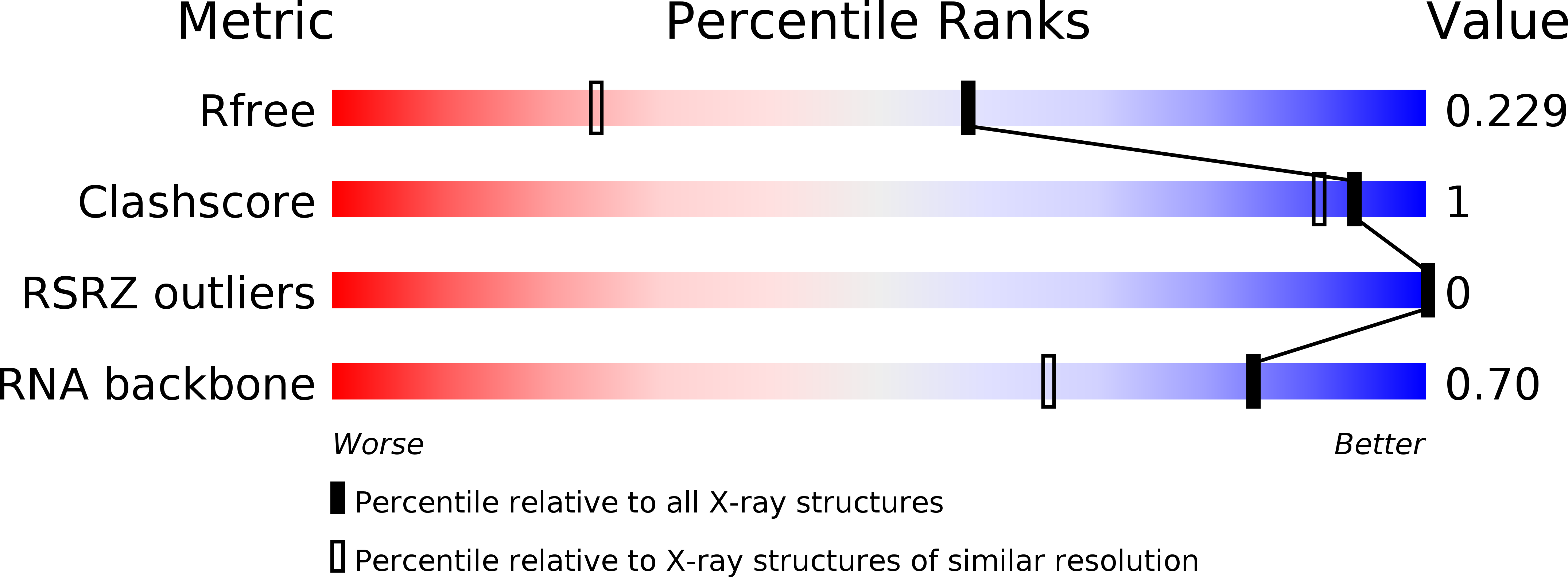
1KD5 image
Entry Detail
PDB ID:
1KD5
Keywords:
Title:
The Crystal Structure of r(GGUCACAGCCC)2 metal free form
Biological Source:
Source Organism:
PDB Version:
Deposition Date:
2001-11-12
Release Date:
2003-03-04
Method Details:
Experimental Method:
Resolution:
1.58 Å
R-Value Free:
0.22
R-Value Work:
0.17
R-Value Observed:
0.17
Space Group:
P 43
Macromolecular Entities
Polymer Type:polyribonucleotide
Description:5'-R(*GP*GP*UP*CP*AP*CP*AP*GP*CP*CP*C)-3'
Chain IDs:A, B
Chain Length:11
Number of Molecules:2
Biological Source:
Primary Citation
Crystal structures of r(GGUCACAGCCC)2.
Acta Crystallogr.,Sect.D 59 423 432 (2003)
PMID: 12595698 DOI: 10.1107/S0907444902021911

Abstact

Crystals of small RNAs, which regularly diffract to very high resolution, can often be readily obtained. Unfortunately, for some RNAs the conformations adopted in the crystalline form are different from those found in solution. For example, short RNAs that form hairpins in solution virtually never crystallize thus; rather, they form duplexes. Nevertheless, these unintended structures have contributed greatly to the understanding of RNA structure. In a similar occurrence, the homodimer r(GGUCACAGCCC)(2) has been crystallized from an 11-mer/12-mer heteroduplex, r(GGCUGAAGUCCG)/r(GGUCACAGCCC). This surprising phenomenon was observed under a variety of crystallization conditions. The structure of the homoduplex was determined from crystals that differed in the precipitant used and the type of metal present. In all cases, the resulting homoduplexes contain ten base pairings, of which the central six are non-canonical pairings. In two of the variants, ordered metal-binding sites were observed: two equivalent octacoordinate Tl(+) sites in one and two equivalent nanocoordinate Ba(2+) sites in another.

Legend

Protein

Chemical

Disease

Primary Citation of related structures