Deposition Date
2001-11-07
Release Date
2002-11-13
Last Version Date
2024-02-07
Entry Detail
PDB ID:
1KBY
Keywords:
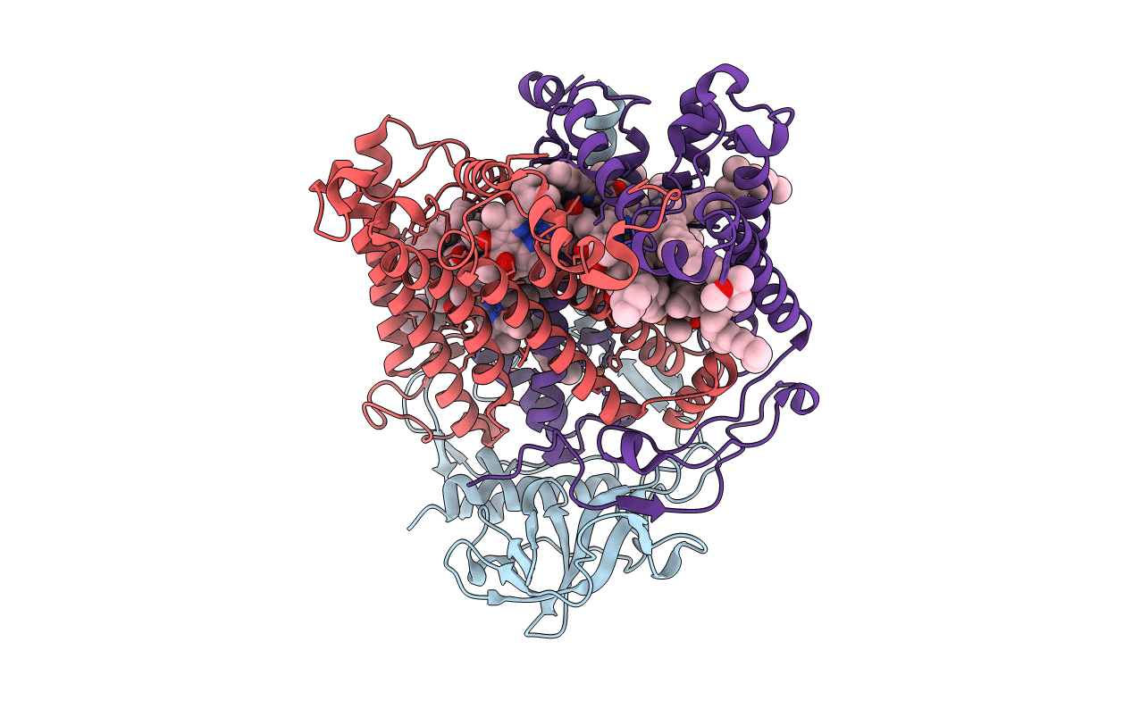
Title:
Structure of Photosynthetic Reaction Center with bacteriochlorophyll-bacteriopheophytin heterodimer
Biological Source:
Source Organism(s):
Rhodobacter sphaeroides (Taxon ID: 1063)
Expression System(s):
Method Details:
Experimental Method:
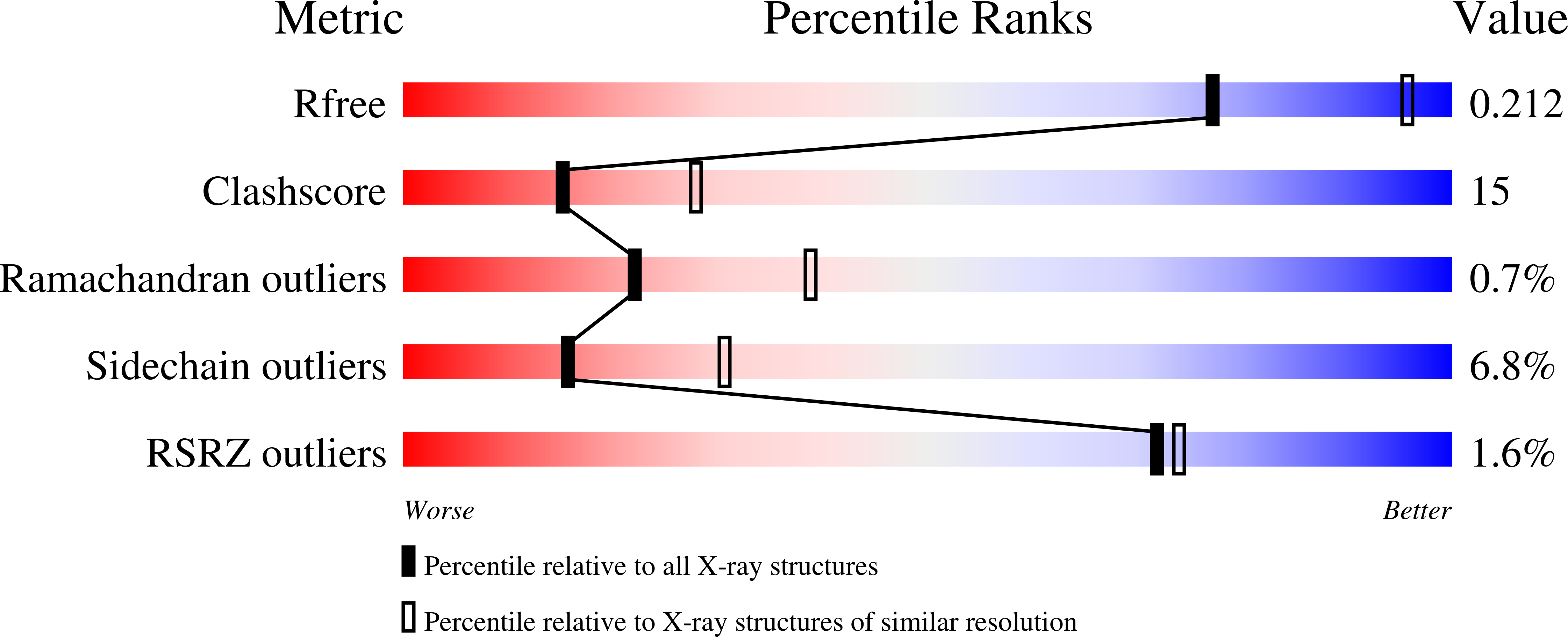
Resolution:
2.50 Å
R-Value Free:
0.22
R-Value Work:
0.19
R-Value Observed:
0.19
Space Group:
P 31 2 1