Deposition Date
1994-07-13
Release Date
1994-12-20
Last Version Date
2024-11-06
Entry Detail
PDB ID:
1KBA
Keywords:
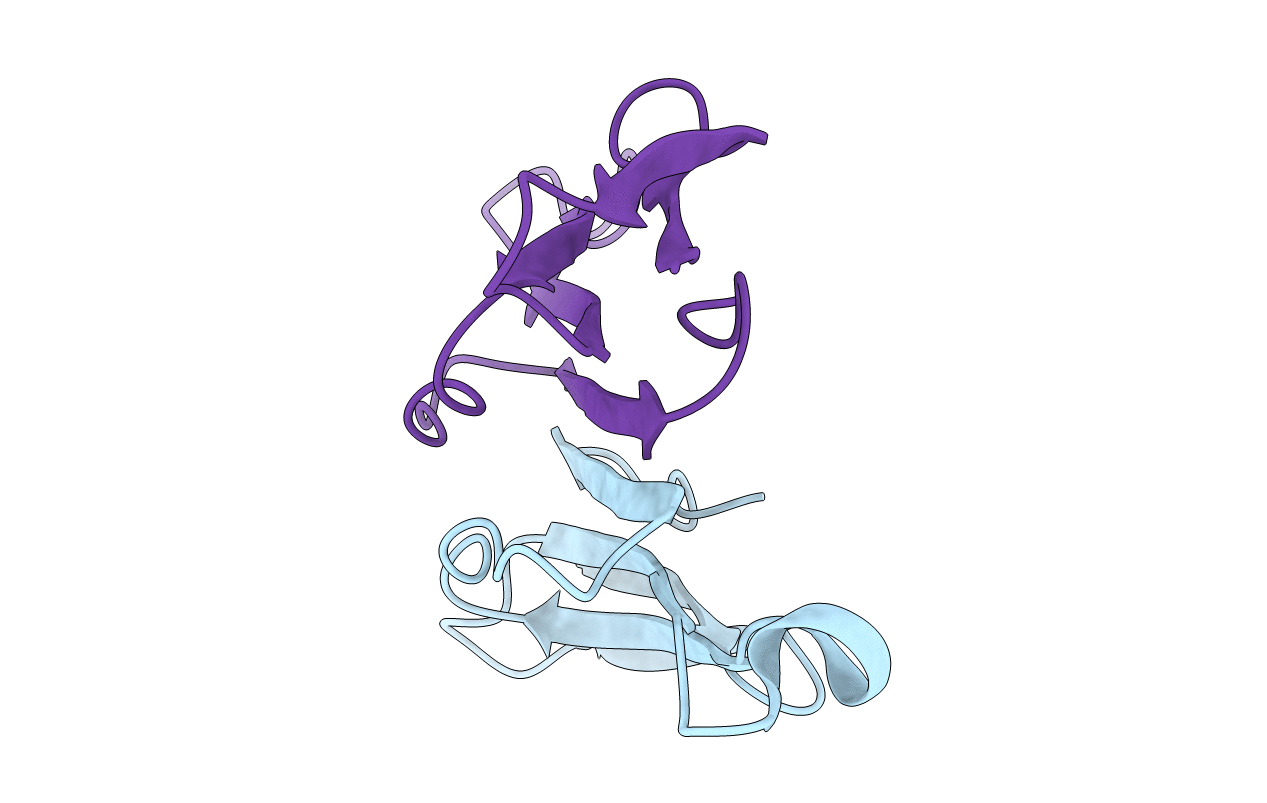
Title:
CRYSTAL STRUCTURE OF KAPPA-BUNGAROTOXIN AT 2.3-ANGSTROM RESOLUTION
Biological Source:
Source Organism(s):
Bungarus multicinctus (Taxon ID: 8616)
Method Details:
Experimental Method:
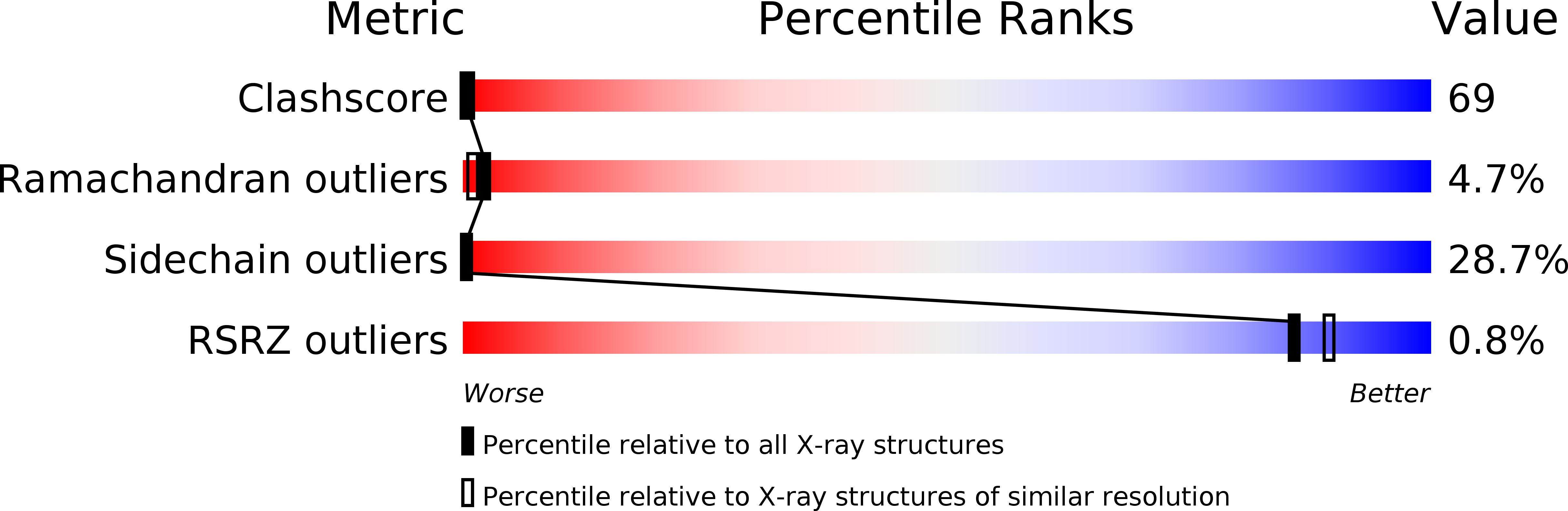
Resolution:
2.30 Å
R-Value Work:
0.19
R-Value Observed:
0.19
Space Group:
P 6