1KB0 image
Deposition Date 2001-11-05
Release Date 2001-12-28
Last Version Date 2024-11-20
Entry Detail
PDB ID:
1KB0
Keywords:
Title:
Crystal Structure of Quinohemoprotein Alcohol Dehydrogenase from Comamonas testosteroni
Biological Source:
Source Organism:
Method Details:
Experimental Method:
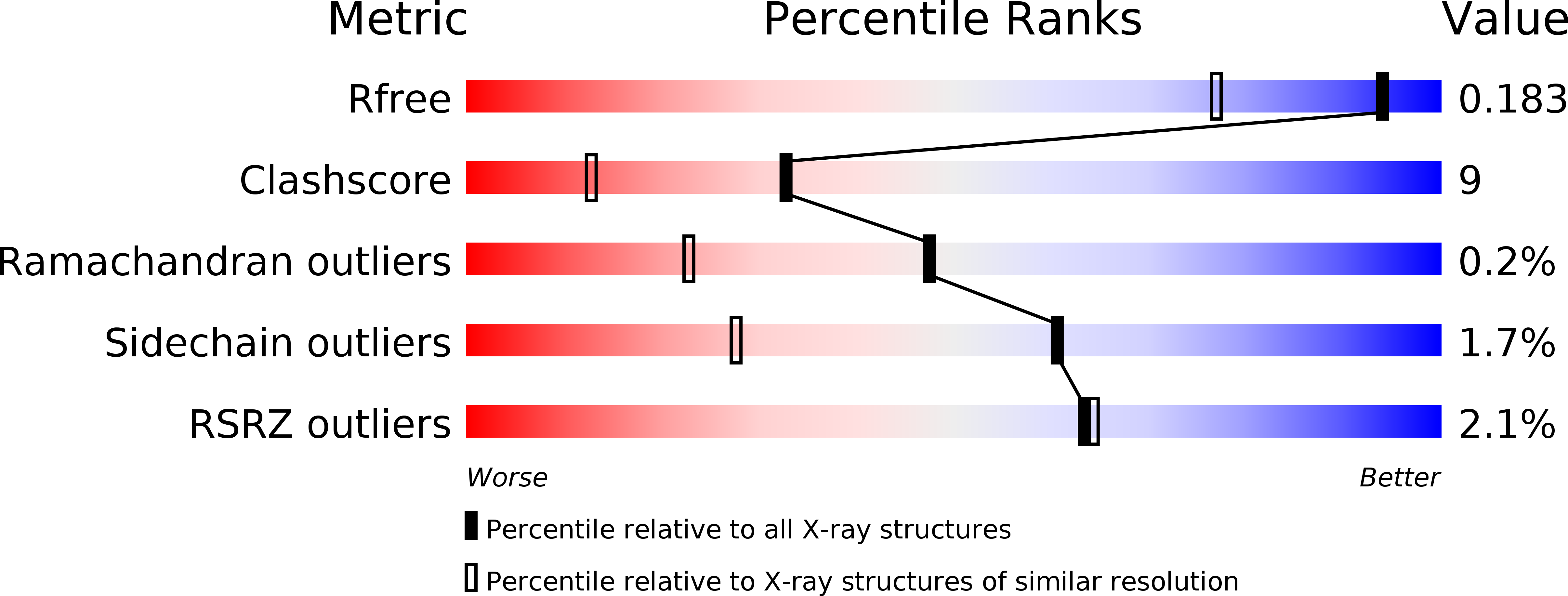
Resolution:
1.44 Å
R-Value Free:
0.18
R-Value Work:
0.16
R-Value Observed:
0.16
Space Group:
C 1 2 1
Macromolecular Entities
Structures with similar UniProt ID
Protein Blast
Polymer Type:polypeptide(L)
Molecule:quinohemoprotein alcohol dehydrogenase
Gene (Uniprot):qheDH
Chain IDs:A
Chain Length:677
Number of Molecules:1
Biological Source:Comamonas testosteroni
Modified Residue
Compound ID Chain ID Parent Comp ID Details 2D Image
TRO A TRP 2-HYDROXY-TRYPTOPHAN
Primary Citation
Crystal structure of quinohemoprotein alcohol dehydrogenase from Comamonas testosteroni: structural basis for substrate oxidation and electron transfer.
J.Biol.Chem. 277 3727 3732 (2002)
PMID: 11714714 DOI: 10.1074/jbc.M109403200

Abstact

Quinoprotein alcohol dehydrogenases are redox enzymes that participate in distinctive catabolic pathways that enable bacteria to grow on various alcohols as the sole source of carbon and energy. The x-ray structure of the quinohemoprotein alcohol dehydrogenase from Comamonas testosteroni has been determined at 1.44 A resolution. It comprises two domains. The N-terminal domain has a beta-propeller fold and binds one pyrroloquinoline quinone cofactor and one calcium ion in the active site. A tetrahydrofuran-2-carboxylic acid molecule is present in the substrate-binding cleft. The position of this oxidation product provides valuable information on the amino acid residues involved in the reaction mechanism and their function. The C-terminal domain is an alpha-helical type I cytochrome c with His(608) and Met(647) as heme-iron ligands. This is the first reported structure of an electron transfer system between a quinoprotein alcohol dehydrogenase and cytochrome c. The shortest distance between pyrroloquinoline quinone and heme c is 12.9 A, one of the longest physiological edge-to-edge distances yet determined between two redox centers. A highly unusual disulfide bond between two adjacent cysteines bridges the redox centers. It appears essential for electron transfer. A water channel delineates a possible pathway for proton transfer from the active site to the solvent.

Legend

Protein

Chemical

Disease

Primary Citation of related structures