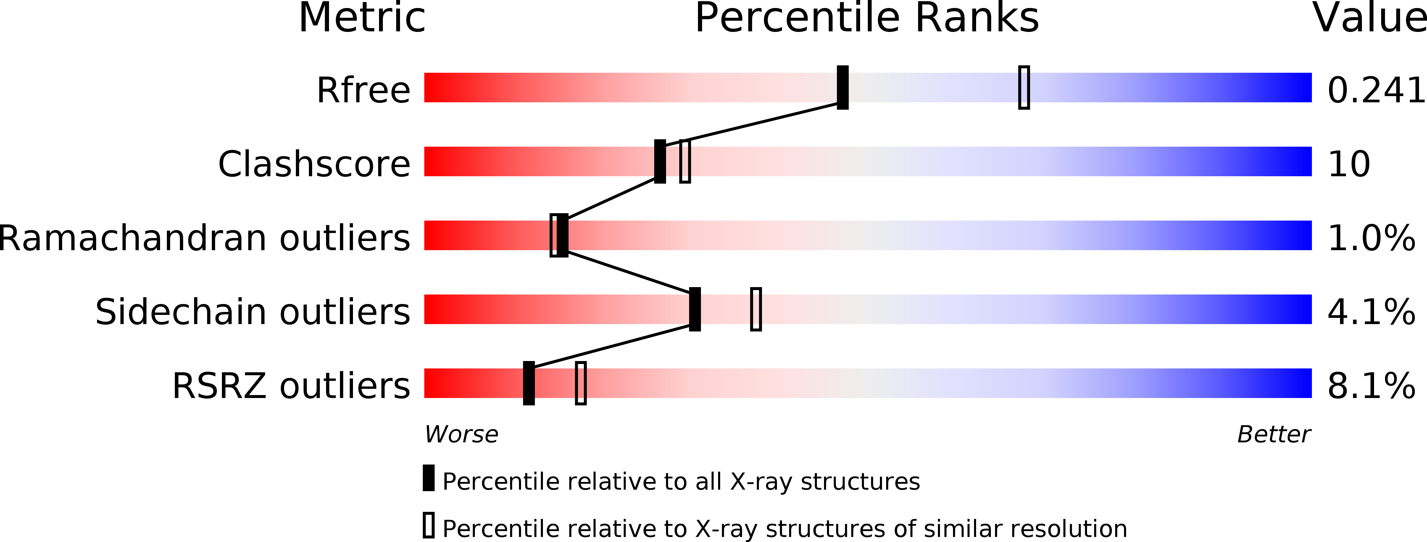
1KAV image
Deposition Date 2001-11-03
Release Date 2002-06-19
Last Version Date 2023-08-16
Entry Detail
PDB ID:
1KAV
Keywords:
Title:
Human Tyrosine Phosphatase 1B Complexed with an Inhibitor
Biological Source:
Source Organism:
Homo sapiens (Taxon ID: 9606)
Host Organism:
Method Details:
Experimental Method:
Resolution:
2.35 Å
R-Value Free:
0.25
R-Value Work:
0.20
R-Value Observed:
0.20
Space Group:
P 31 2 1
Macromolecular Entities
Polymer Type:polypeptide(L)
Molecule:PROTEIN-TYROSINE PHOSPHATASE, NON-RECEPTOR TYPE 1
Gene (Uniprot):PTPN1
Chain IDs:A
Chain Length:298
Number of Molecules:1
Biological Source:Homo sapiens
Ligand Molecules
Primary Citation
Structure of protein tyrosine phosphatase 1B in complex with inhibitors bearing two phosphotyrosine mimetics.
J.Med.Chem. 44 4584 4594 (2001)
PMID: 11741477 DOI: 10.1021/jm010266w

Abstact

Protein tyrosine phosphatases (PTPases) are signal-transducing enzymes that dephosphorylate intracellular proteins that have phosphorylated tyrosine residues. It has been demonstrated that protein tyrosine phosphatase 1B (PTP1B) is an attractive therapeutic target because of its involvement in regulating insulin sensitivity (Elcheby et al. Science 1999, 283, 1544-1548). The identification of a second binding site in PTP1B (Puius et al., Proc. Natl. Acad. Sci. U.S.A.1997, 94, 13420-13425) suggests a new strategy for inhibitor design, where appropriate compounds may be made to simultaneously occupy both binding sites to gain much higher affinity and selectivity. To test this hypothesis and gain further insights into the structural basis of inhibitor binding, we have determined the crystal structure of PTP1B complexed with two non-peptidyl inhibitors, 4 and 5, both of which contain two aryl difluoromethylenephosphonic acid groups, a nonhydrolyzable phosphate mimetic. The structures were determined and refined to 2.35 and 2.50 A resolution, respectively. Although one of the inhibitors seems to have satisfied the perceived requirement for dual binding, it did not bind both the active site and the adjacent noncatalytic binding site as expected. The second or distal phosphonate group instead extends into the solvent and makes water-mediated interactions with Arg-47. The selectivity of the more potent of these two inhibitors, as well as four other inhibitors bearing two such phosphate mimetics for PTP1B versus seven other PTPases, was examined. In general, selectivity was modest to good when compared to PTPases Cdc25a, PTPmeg-1, PTPbeta, and CD45. However, selectivity was generally poor when compared to other PTPases such as SHP-1, SHP-2, and especially TCPTP, for which almost no selectivity was found. The implications these results have concerning the utility of dual-binding inhibitors are discussed.

Legend

Protein

Chemical

Disease

Primary Citation of related structures