Deposition Date
2001-10-29
Release Date
2002-04-10
Last Version Date
2023-08-16
Entry Detail
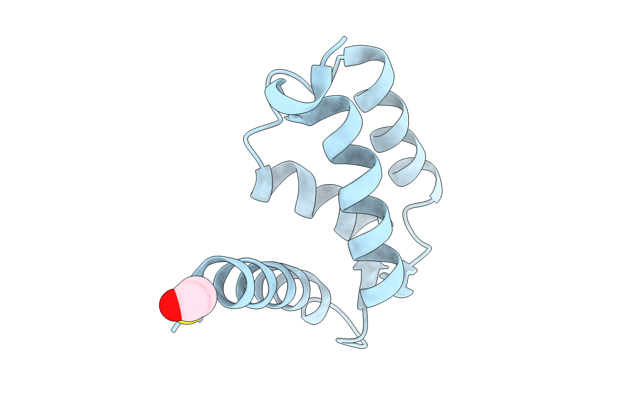
PDB ID:
1K9P
Keywords:
Title:
CRYSTAL STRUCTURE OF CALCIUM FREE (OR APO) HUMAN S100A6
Biological Source:
Source Organism(s):
Homo sapiens (Taxon ID: 9606)
Expression System(s):
Method Details:
Experimental Method:
Resolution:
1.90 Å
R-Value Free:
0.18
R-Value Work:
0.17
R-Value Observed:
0.17
Space Group:
C 2 2 21