Deposition Date
2001-10-25
Release Date
2001-12-31
Last Version Date
2024-02-07
Entry Detail
PDB ID:
1K8W
Keywords:
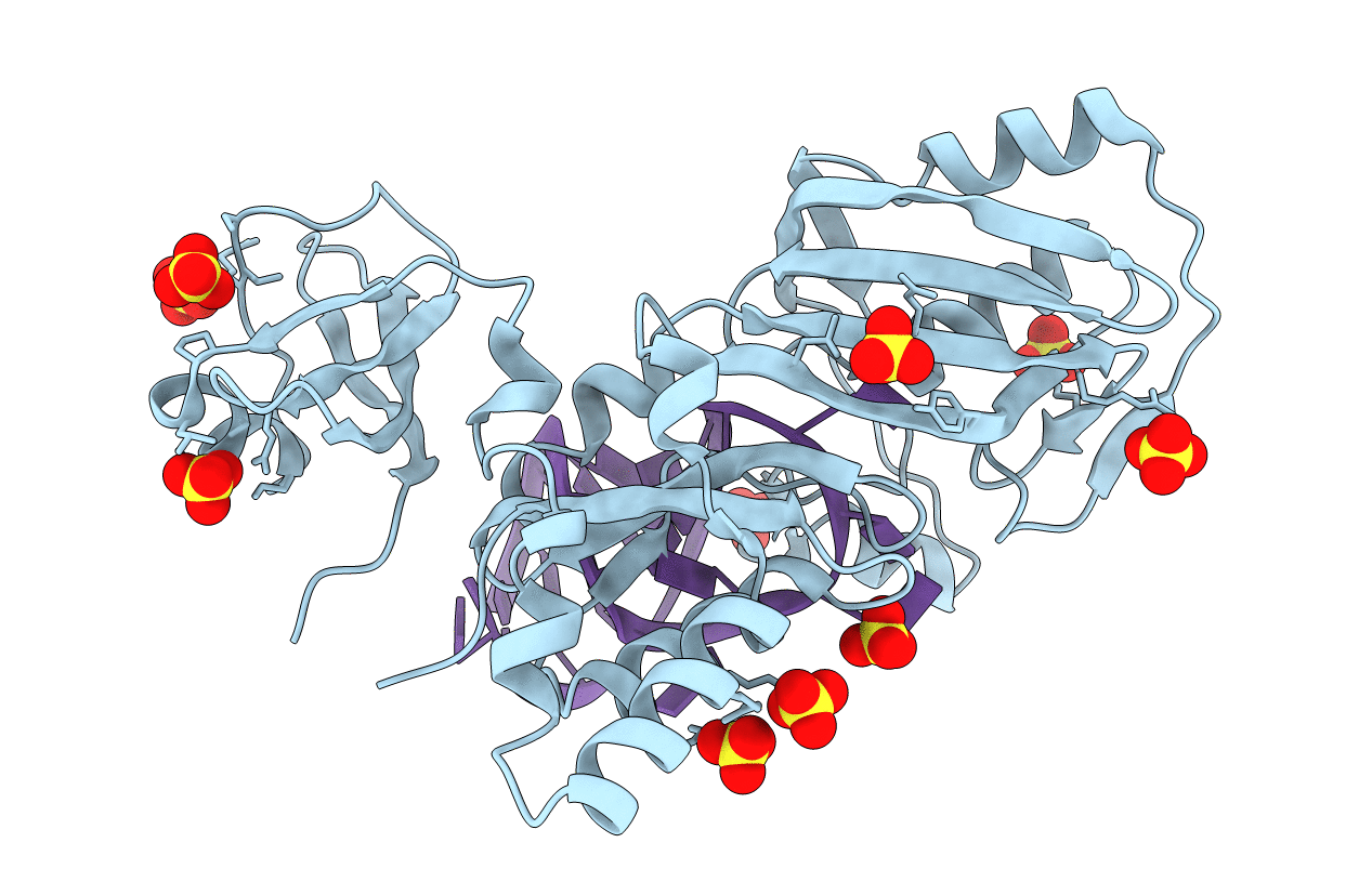
Title:
Crystal structure of the E. coli pseudouridine synthase TruB bound to a T stem-loop RNA
Biological Source:
Source Organism(s):
Escherichia coli (Taxon ID: 562)
Expression System(s):
Method Details:
Experimental Method:
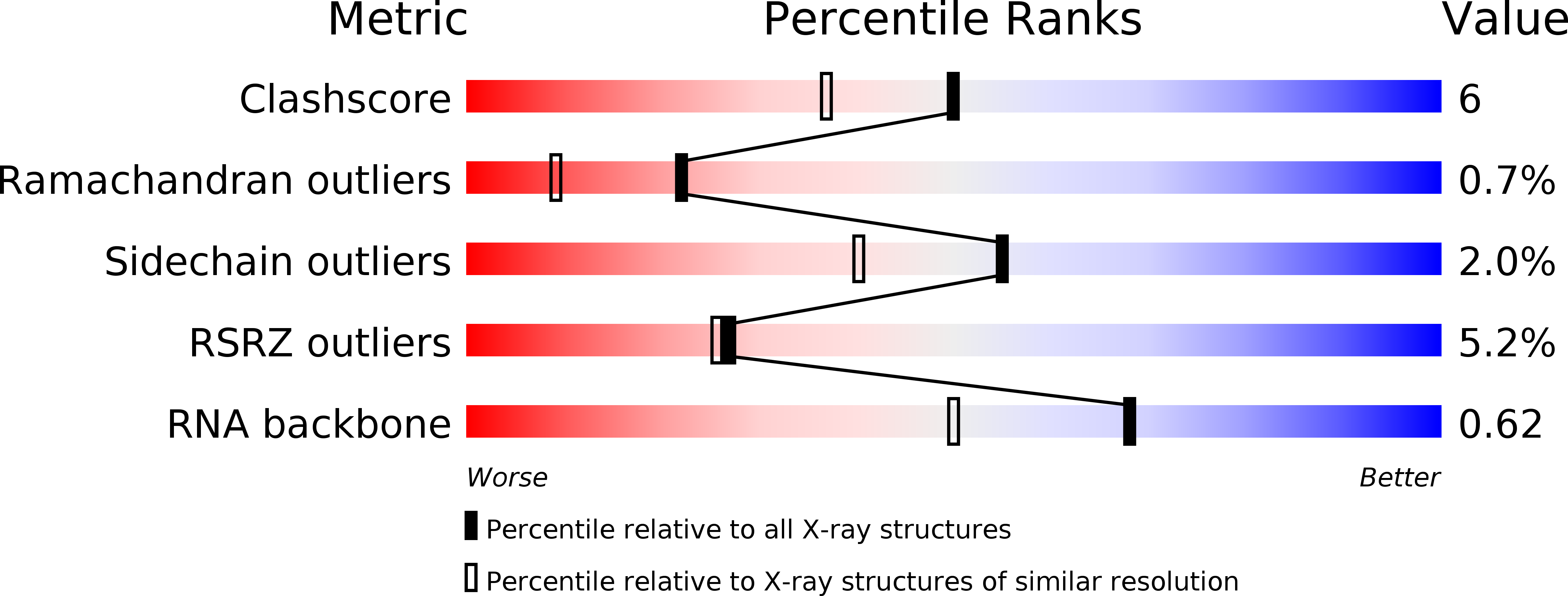
Resolution:
1.85 Å
R-Value Free:
0.21
R-Value Work:
0.18
R-Value Observed:
0.18
Space Group:
C 1 2 1