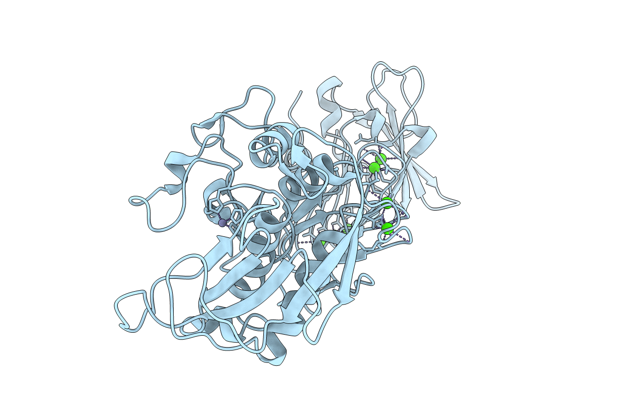
1K7Q image
Deposition Date 2001-10-20
Release Date 2002-10-20
Last Version Date 2024-02-07
Entry Detail
PDB ID:
1K7Q
Keywords:
Title:
PrtC from Erwinia chrysanthemi: E189A mutant
Biological Source:
Source Organism:
Host Organism:
Method Details:
Experimental Method:
Resolution:
1.80 Å
R-Value Free:
0.21
R-Value Work:
0.19
R-Value Observed:
0.20
Space Group:
P 31 2 1
Macromolecular Entities
Structures with similar UniProt ID
Protein Blast
Polymer Type:polypeptide(L)
Molecule:SECRETED PROTEASE C
Gene (Uniprot):prtC
Mutagens:E189A
Chain IDs:A
Chain Length:479
Number of Molecules:1
Biological Source:Erwinia chrysanthemi
Primary Citation
Protease C of Erwinia chrysanthemi: The Crystal Structure and Role of Amino Acids Y228 and E189
J.Mol.Biol. 314 187 193 (2001)
PMID: 11718553 DOI: 10.1006/jmbi.2001.5124

Abstact

PrtC, a metallo-protease secreted by Erwinia chrysanthemi, is a member of the serralysin family and hence belongs to the metzincin superfamily. While the crystal structures of representatives of all metzincin subfamilies have been elucidated in the past, there is still some controversy about the reaction mechanism and the role of certain characteristic amino acids in the active centre. In this study, we probed the role of Tyr228 and Glu189 by site-directed mutagenesis and X-ray crystallography. There is evidence that these residues participate in catalysis, although conflicting hypotheses have been proposed. The crystal structures of wild-type and mutants have been refined to an R(free) of about 0.20 at resolutions of 2.0 A or better. Exchange of Glu189 versus alanine reduces the catalytic efficiency to less than 0.5 % using resorufin casein as substrate and to about 3 % using an assay utilising the thiol ester Ac-Pro-Leu-Gly-[(S)Leu]-Leu-Gly-OEt. The drop in activity is caused by a reduction in k(cat) while the K(M) values are virtually the same. In the resorufin casein assay, the mutant Y228F shows about 3 % of the wild-type activity and in the thiol ester assay this increases to about 56 %. In the latter case, the K(M) value of the mutant is increased from 5.3 mM to 9.0 mM with only little reduction in k(cat). The different behaviour of this mutant with respect to the two substrates can be explained by a switch in the rate-determining step during catalysis. The study presented here provides clear evidence that Glu189 of the HEXXHXXGXXH motif is the catalytic base, while Tyr228 is more likely involved in substrate binding and the stabilisation of the tetrahedral transition state.

Legend

Protein

Chemical

Disease

Primary Citation of related structures