1K7C image
Deposition Date 2001-10-19
Release Date 2001-12-28
Last Version Date 2024-10-23
Entry Detail
PDB ID:
1K7C
Keywords:
Title:
Rhamnogalacturonan acetylesterase with seven N-linked carbohydrate residues distributed at two N-glycosylation sites refined at 1.12 A resolution
Biological Source:
Source Organism:
Host Organism:
Method Details:
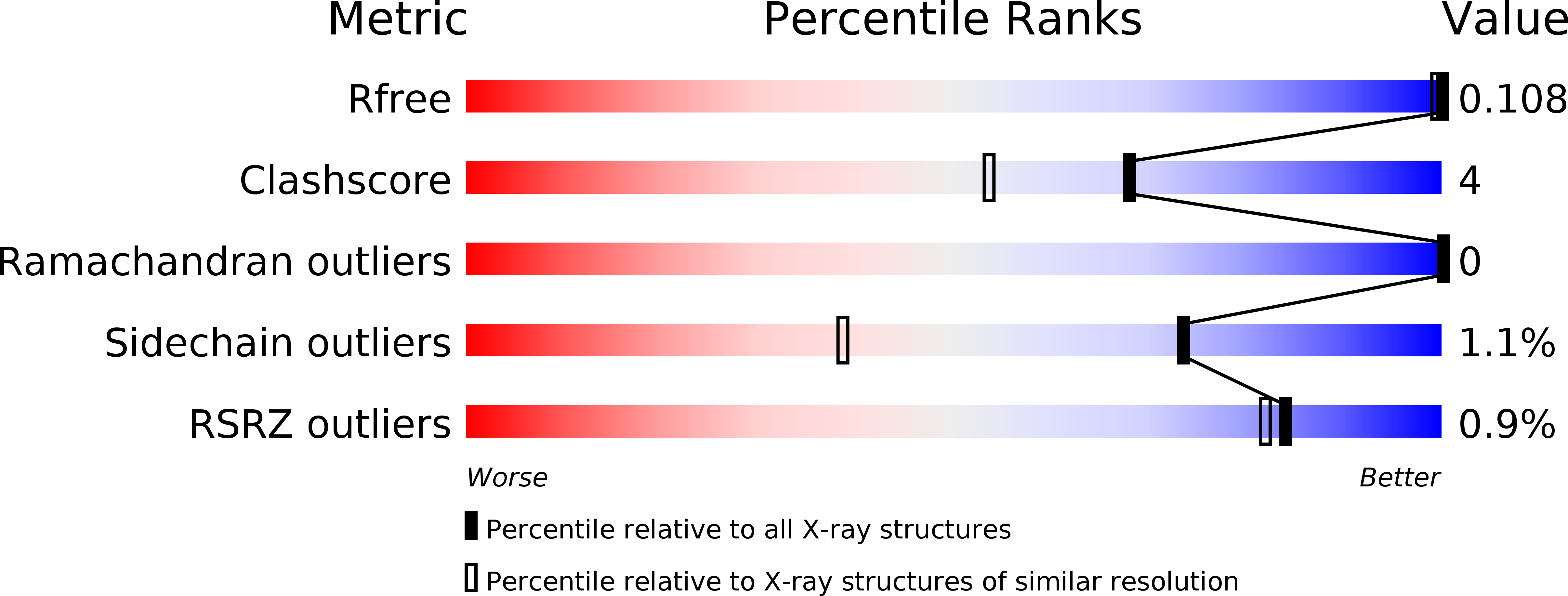
Experimental Method:
Resolution:
1.12 Å
R-Value Free:
0.13
R-Value Work:
0.10
R-Value Observed:
0.10
Space Group:
P 21 21 21
Macromolecular Entities
Structures with similar UniProt ID
Protein Blast
Polymer Type:polypeptide(L)
Molecule:rhamnogalacturonan acetylesterase
Chain IDs:A
Chain Length:233
Number of Molecules:1
Biological Source:Aspergillus aculeatus
Modified Residue
Compound ID Chain ID Parent Comp ID Details 2D Image
ASN A ASN GLYCOSYLATION SITE
Primary Citation
A branched N-linked glycan at atomic resolution in the 1.12 A structure of rhamnogalacturonan acetylesterase.
Acta Crystallogr.,Sect.D 58 111 119 (2002)
PMID: 11752785 DOI: 10.1107/S0907444901018479

Abstact

The crystal structure of the glycoprotein rhamnogalacturonan acetylesterase from Aspergillus aculeatus has been refined to a resolution of 1.12 A using synchrotron data collected at 263 K. Both of the two putative N-glycosylation sites at Asn104 and Asn182 are glycosylated and, owing to crystal contacts, the glycan structure at Asn182 is exceptionally well defined in the electron-density maps, showing the six-carbohydrate structure Manalpha1-6(Manalpha1-3)Manalpha1-6Manbeta1-4GlcNAcbeta1-4GlcNAcbeta-Asn182. Equivalent carbohydrate residues were restrained to have similar geometries, but were refined without target values. The refined bond lengths and angles were compared with the values obtained from small-molecule studies that form the basis for the dictionaries used for glycoprotein refinement.

Legend

Protein

Chemical

Disease

Primary Citation of related structures