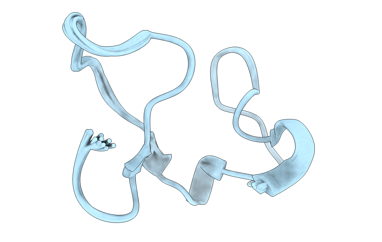
1K7B image
Deposition Date 2001-10-18
Release Date 2001-12-19
Last Version Date 2024-11-20
Entry Detail
PDB ID:
1K7B
Title:
NMR Solution Structure of sTva47, the Viral-Binding Domain of Tva
Biological Source:
Source Organism:
Host Organism:
Method Details:
Experimental Method:
Conformers Calculated:
100
Conformers Submitted:
20
Selection Criteria:
structures with the lowest energy
Macromolecular Entities
Polymer Type:polypeptide(L)
Molecule:SUBGROUP A ROUS SARCOMA VIRUS RECEPTOR PG800 AND PG950
Chain IDs:A
Chain Length:47
Number of Molecules:1
Biological Source:Coturnix coturnix
Primary Citation
The solution structure of the viral binding domain of Tva, the cellular receptor for subgroup A avian leukosis and sarcoma virus.
FEBS Lett. 509 161 168 (2001)
PMID: 11768384 DOI: 10.1016/S0014-5793(01)03086-1

Abstact

The cellular receptor for subgroup A avian leukosis and sarcoma virus (ALSV-A) is Tva, which contains a motif related to repeats in the low density lipoprotein receptor (LDLR) ligand binding repeat (LBr) and which is necessary for viral entry. As observed with LBr repeats of LDLR, the 47 residue LBr domain of Tva (sTva47) requires calcium during oxidative folding to form the correct disulfide bonds, and calcium enhances the structure of correctly oxidized sTva47, as well as its ability to bind the viral envelope protein (Env). However, solution nuclear magnetic resonance studies indicate that, even in the presence of excess calcium, sTva47 exists in an ensemble of conformations. Nonetheless, as reported here, the structure of the predominant sTva47 solution conformer closely resembles that of other LBr repeats, with identical S-S binding topology and octahedral calcium coordination. The location of W48 and other critical residues on the surface suggests a region of the molecule necessary for Env binding and to mediate post-binding events important for ALSV-A cell entry.

Legend

Protein

Chemical

Disease

Primary Citation of related structures