
Deposition Date
2001-10-18
Release Date
2003-07-22
Last Version Date
2023-08-16
Entry Detail
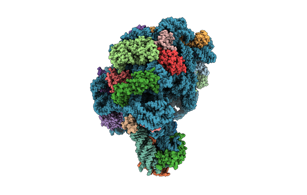
PDB ID:
1K73
Keywords:
Title:
Co-crystal Structure of Anisomycin Bound to the 50S Ribosomal Subunit
Biological Source:
Source Organism(s):
Haloarcula marismortui (Taxon ID: 2238)
Method Details:
Experimental Method:
Resolution:
3.01 Å
R-Value Free:
0.24
R-Value Work:
0.21
R-Value Observed:
0.21
Space Group:
C 2 2 21
