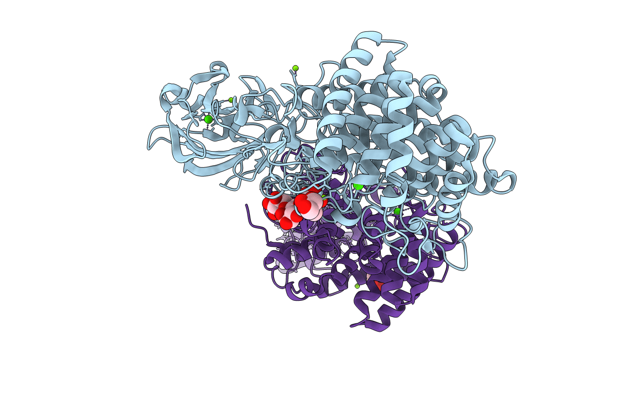
1K72 image
Deposition Date 2001-10-18
Release Date 2003-07-15
Last Version Date 2023-08-16
Entry Detail
PDB ID:
1K72
Keywords:
Title:
The X-ray Crystal Structure Of Cel9G Complexed With cellotriose
Biological Source:
Source Organism:
Host Organism:
Method Details:
Experimental Method:
Resolution:
1.80 Å
R-Value Free:
0.19
R-Value Work:
0.16
R-Value Observed:
0.16
Space Group:
P 1
Macromolecular Entities
Polymer Type:polypeptide(L)
Molecule:Endoglucanase 9G
Gene (Uniprot):celCCG
Chain IDs:A, B
Chain Length:614
Number of Molecules:2
Biological Source:Clostridium cellulolyticum
Modified Residue
Compound ID Chain ID Parent Comp ID Details 2D Image
SCH A CYS S-METHYL-THIO-CYSTEINE
Peptide-like Molecules
PRD_900005
Primary Citation
X-Ray Crystal Structure of the Multidomain Endoglucanase Cel9G from Clostridium cellulolyticum Complexed with Natural and Synthetic Cello-Oligosaccharides
J.BACTERIOL. 185 4127 4135 (2003)
PMID: 12837787 DOI: 10.1128/JB.185.14.4127-4135.2003

Abstact

Complete cellulose degradation is the first step in the use of biomass as a source of renewable energy. To this end, the engineering of novel cellulase activity, the activity responsible for the hydrolysis of the beta-1,4-glycosidic bonds in cellulose, is a topic of great interest. The high-resolution X-ray crystal structure of a multidomain endoglucanase from Clostridium cellulolyticum has been determined at a 1.6-A resolution. The endoglucanase, Cel9G, is comprised of a family 9 catalytic domain attached to a family III(c) cellulose-binding domain. The two domains together form a flat platform onto which crystalline cellulose is suggested to bind and be fed into the active-site cleft for endolytic hydrolysis. To further dissect the structural basis of cellulose binding and hydrolysis, the structures of Cel9G in the presence of cellobiose, cellotriose, and a DP-10 thio-oligosaccharide inhibitor were resolved at resolutions of 1.7, 1.8, and 1.9 A, respectively.

Legend

Protein

Chemical

Disease

Primary Citation of related structures