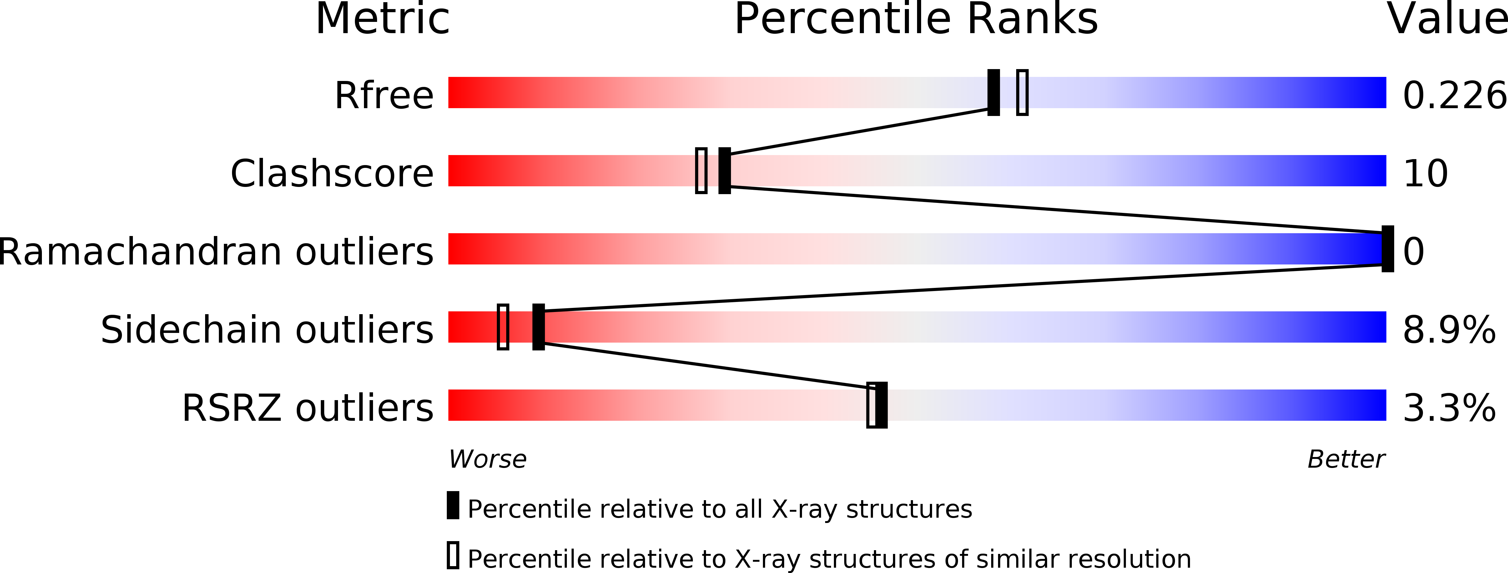
1K6Z image
Deposition Date 2001-10-17
Release Date 2001-10-31
Last Version Date 2024-11-20
Entry Detail
PDB ID:
1K6Z
Keywords:
Title:
Crystal Structure of the Yersinia Secretion Chaperone SycE
Biological Source:
Source Organism:
Yersinia pestis (Taxon ID: 632)
Method Details:
Experimental Method:
Resolution:
2.00 Å
R-Value Free:
0.24
R-Value Work:
0.19
Space Group:
P 1 21 1
Macromolecular Entities
Structures with similar UniProt ID
Protein Blast
Polymer Type:polypeptide(L)
Molecule:Type III secretion chaperone SycE
Gene (Uniprot):yerA
Chain IDs:A, B
Chain Length:141
Number of Molecules:2
Biological Source:Yersinia pestis
Modified Residue
Compound ID Chain ID Parent Comp ID Details 2D Image
MSE A MET SELENOMETHIONINE
Ligand Molecules
Primary Citation
Three-dimensional structure of the type III secretion chaperone SycE from Yersinia pestis.
Acta Crystallogr.,Sect.D 58 398 406 (2002)
PMID: 11856824 DOI: 10.1107/S090744490200015X

Abstact

Many bacterial pathogens utilize a type III (contact-dependent) secretion system to inject cytotoxic effector proteins directly into host cells. This ingenious mechanism, designed for both bacterial offense and defense, has been studied most extensively in Yersinia spp. To be exported efficiently, at least three of the effectors (YopE, YopH and YopT) and several other proteins that transit the type III secretion pathway in Yersinia (YopN, YopD and YopB) must first form transient complexes with cognate-specific Yop chaperone (Syc) proteins. The cytotoxic effector YopE, a selective activator of mammalian Rho-family GTPases, associates with SycE. Here, the structure of Y. pestis SycE at 1.95A resolution is reported. SycE possesses a novel fold with an unusual dimerization motif and an intriguing basic cavity located on the dyad axis of the dimer that may participate in its interaction with YopE.

Legend

Protein

Chemical

Disease

Primary Citation of related structures