1K66 image
Deposition Date 2001-10-15
Release Date 2003-12-16
Last Version Date 2024-02-07
Entry Detail
PDB ID:
1K66
Title:
Crystal Structure of the Cyanobacterial Phytochrome Response Regulator, RcpB
Biological Source:
Source Organism:
Host Organism:
Method Details:
Experimental Method:
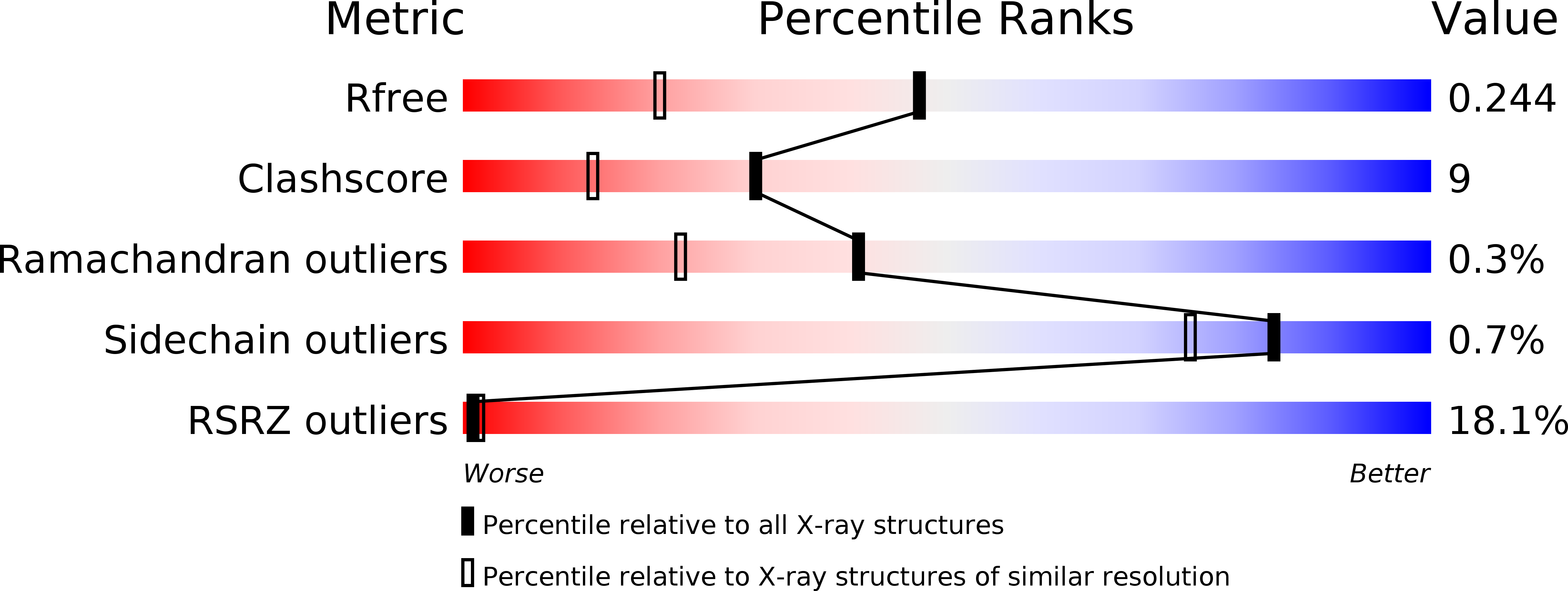
Resolution:
1.75 Å
R-Value Free:
0.25
R-Value Work:
0.21
R-Value Observed:
0.21
Space Group:
P 41 21 2
Macromolecular Entities
Structures with similar UniProt ID
Protein Blast
Polymer Type:polypeptide(L)
Molecule:Phytochrome Response Regulator RcpB
Gene (Uniprot):rcpB
Chain IDs:A, B
Chain Length:149
Number of Molecules:2
Biological Source:Tolypothrix sp. PCC 7601
Ligand Molecules
Primary Citation
Crystal structures of two cyanobacterial response regulators in apo- and phosphorylated form reveal a novel dimerization motif of phytochrome-associated response regulators
Biophys.J. 87 476 487 (2004)
PMID: 15240481 DOI: 10.1529/biophysj.103.033696

Abstact

The structures of two response regulators (RRs) from the cyanobacterium Calothrix PCC7601, RcpA and RcpB, were solved to 1.9- and 1.75-A resolution, respectively. RcpA was found in phosphorylated and RcpB in nonphosphorylated form. Both RRs are members of phytochrome-associated, light-sensing two-component signal transduction pathways, based on histidine kinase-mediated receptor autophosphorylation and phosphorelay to a RR. Despite the overall folding similarity to CheY-type RRs ((beta/alpha)(5)-motif), RcpA and RcpB form homodimers, irrespective of their phosphorylation state, giving insight into a signal transduction putatively different from that of other known RRs. Dimerization is accomplished by a C-terminal extension of the RR polypeptide chain, and the surface formed by H4, beta 5, and H5, which constitute a hydrophobic contact area with distinct interactions between residues of either subunit. Sequence alignments reveal that the identified dimerization motif is archetypal for phytochrome-associated RRs, making them a novel subgroup of CheY-type RRs. The protein structures of RcpA and RcpB are compared to the recently presented protein structure of Rcp1 from Synechocystis.

Legend

Protein

Chemical

Disease

Primary Citation of related structures
Feedback Form
Name
Email
Institute
Feedback