1K62 image
Deposition Date 2001-10-14
Release Date 2002-02-01
Last Version Date 2023-08-16
Entry Detail
PDB ID:
1K62
Keywords:
Title:
Crystal Structure of the Human Argininosuccinate Lyase Q286R Mutant
Biological Source:
Source Organism:
Homo sapiens (Taxon ID: 9606)
Host Organism:
Method Details:
Experimental Method:
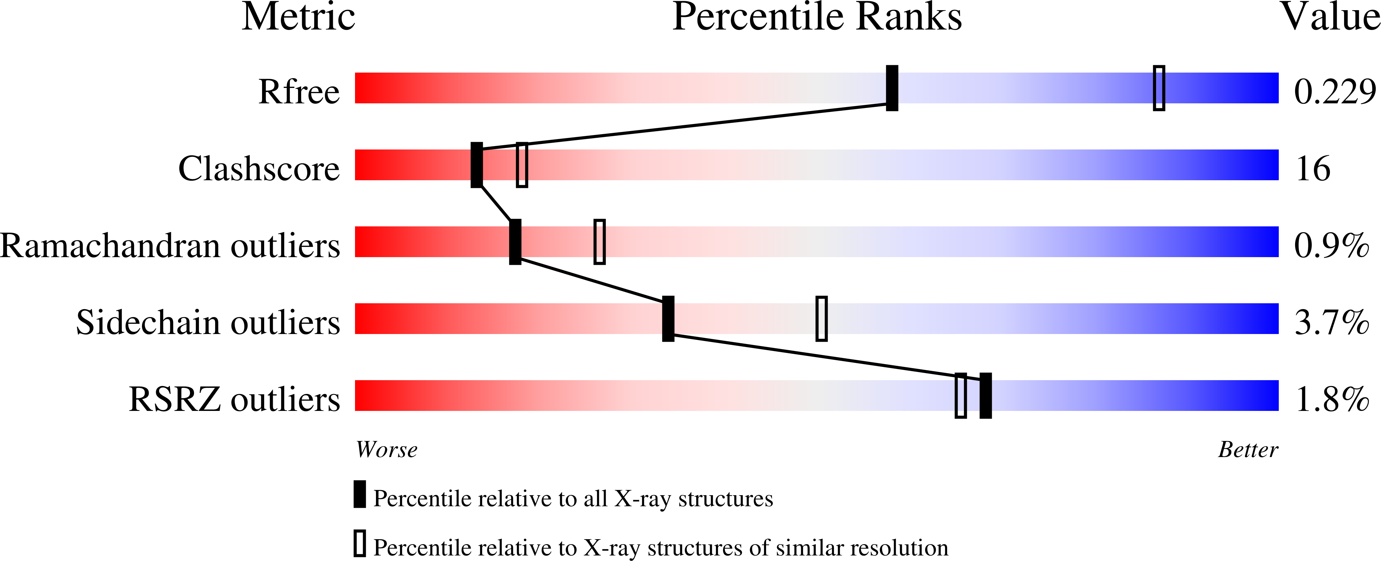
Resolution:
2.65 Å
R-Value Free:
0.22
R-Value Work:
0.17
R-Value Observed:
0.17
Space Group:
P 31 2 1
Macromolecular Entities
Structures with similar UniProt ID
Protein Blast
Polymer Type:polypeptide(L)
Molecule:Argininosuccinate Lyase
Gene (Uniprot):ASL
Mutagens:Q286R
Chain IDs:A, B
Chain Length:464
Number of Molecules:2
Biological Source:Homo sapiens
Primary Citation
Three-dimensional structure of the argininosuccinate lyase frequently complementing allele Q286R.
Biochemistry 40 15570 15580 (2001)
PMID: 11747432 DOI: 10.1021/bi011525m

Abstact

Argininosuccinate lyase (ASL) catalyzes the reversible breakdown of argininosuccinate to arginine and fumarate, a reaction involved in the biosynthesis of arginine in all species and in the production of urea in ureotelic species. In humans, mutations in the enzyme result in the autosomal recessive disorder argininosuccinic aciduria. Intragenic complementation has been demonstrated to occur at the ASL locus, with two distinct classes of ASL-deficient strains having been identified, the frequent and high-activity complementers. The frequent complementers participate in the majority of the complementation events observed and were found to be either homozygous or heterozygous for a glutamine to arginine mutation at residue 286. The three-dimensional structure of the frequently complementing allele Q286R has been determined at 2.65 A resolution. This is the first high-resolution structure of human ASL. Comparison of this structure with the structures of wild-type and mutant duck delta1 and delta2 crystallins suggests that the Q286R mutation may sterically and/or electrostatically hinder a conformational change in the 280's loop (residues 270-290) and domain 3 that is thought to be necessary for catalysis to occur. The comparison also suggests that residues other than R33, F333, and D337 play a role in maintaining the structural integrity of domain 1 and reinforces the suggestion that residues 74-89 require a particular conformation for catalysis. The electron density has enabled the structure of residues 6-18 to be modeled for the first time. Residues 7-9 and 15-18 are in type IV beta-turns and are connected by a loop. The conformation observed is stabilized, in part, by a salt bridge between the side chains of R12 and D18. Although the disease causing mutation R12Q would disrupt this salt bridge, it is unclear why this mutation has such a significant effect on the catalytic activity as residues 1-18 are disordered in all other delta-crystallin structures determined to date.

Legend

Protein

Chemical

Disease

Primary Citation of related structures