1K5U image
Deposition Date 2001-10-12
Release Date 2002-02-27
Last Version Date 2023-08-16
Entry Detail
PDB ID:
1K5U
Title:
Human acidic fibroblast growth factor. 141 amino acid form with amino terminal His tag with His93 replaced by Gly (H93G).
Biological Source:
Source Organism:
Homo sapiens (Taxon ID: 9606)
Host Organism:
Method Details:
Experimental Method:
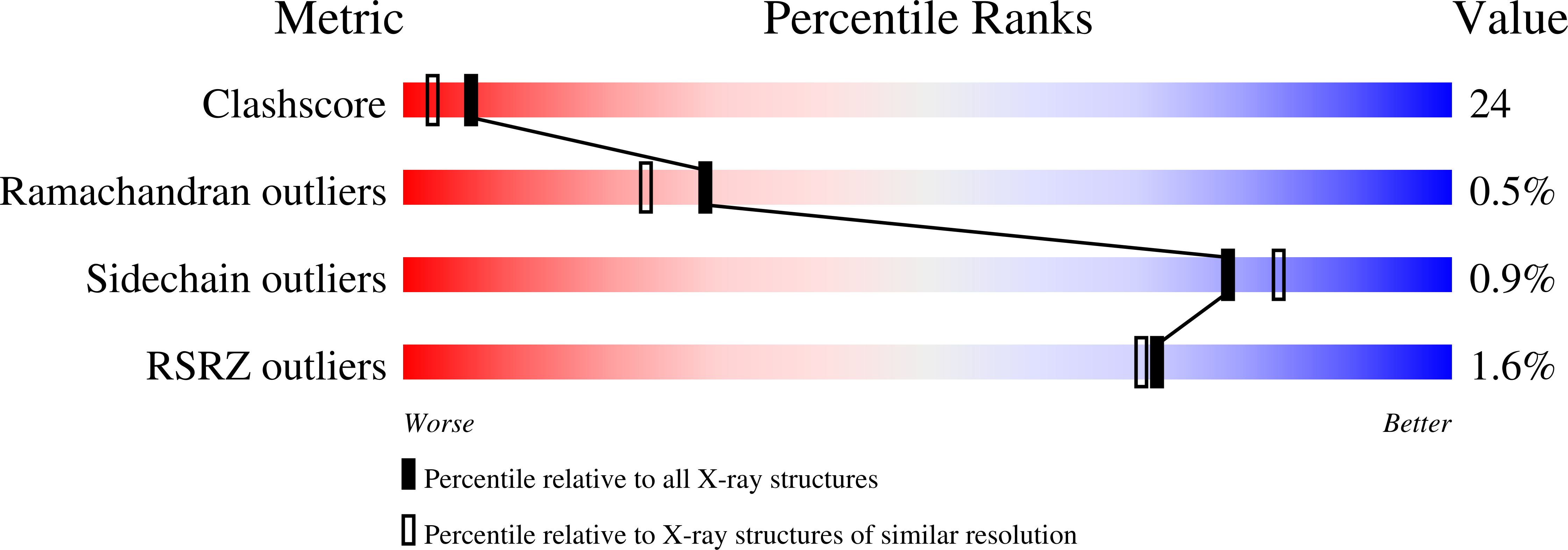
Resolution:
2.00 Å
R-Value Free:
0.29
R-Value Work:
0.21
R-Value Observed:
0.23
Space Group:
P 1 21 1
Macromolecular Entities
Polymer Type:polypeptide(L)
Molecule:Acidic fibroblast growth factor
Gene (Uniprot):FGF1
Mutations:H93G
Chain IDs:A, B, C
Chain Length:146
Number of Molecules:3
Biological Source:Homo sapiens
Ligand Molecules
Primary Citation
Alternative type I and I' turn conformations in the beta8/beta9 beta-hairpin of human acidic fibroblast growth factor.
Protein Sci. 11 459 466 (2002)
PMID: 11847269 DOI: 10.1110/ps.43802

Abstact

Human acidic fibroblast growth factor (FGF-1) has a beta-trefoil structure, one of the fundamental protein superfolds. The X-ray crystal structures of wild-type and various mutant forms of FGF-1 have been solved in five different space groups: C2, C222(1), P2(1) (four molecules/asu), P2(1) (three molecules/asu), and P2(1)2(1)2(1). These structures reveal two characteristically different conformations for the beta8/beta9 beta-hairpin comprising residue positions 90-94. This region in the wild-type FGF-1 structure (P2(1), four molecules/asu), a his-tagged His93-->Gly mutant (P2(1), three molecules/asu) and a his-tagged Asn106-->Gly mutant (P2(1)2(1)2(1)) adopts a 3:5 beta-hairpin known as a type I (1-4) G1 beta-bulge (containing a type I turn). However, a his-tagged form of wild-type FGF-1 (C222(1)) and a his-tagged Leu44-->Phe mutant (C2) adopt a 3:3 beta-hairpin (containing a type I' turn) for this same region. A feature that distinguishes these two types of beta-hairpin structures is the number and location of side chain positions with eclipsed C(beta) and main-chain carbonyl oxygen groups (Psi is equivalent to +60 degrees). The effects of glycine mutations upon stability, at positions within the hairpin, have been used to identify the most likely structure in solution. Type I' turns in the structural data bank are quite rare, and a survey of these turns reveals that a large percentage exhibit crystal contacts within 3.0 A. This suggests that many of the type I' turns in X-ray structures may be adopted due to crystal packing effects.

Legend

Protein

Chemical

Disease

Primary Citation of related structures