Abstact
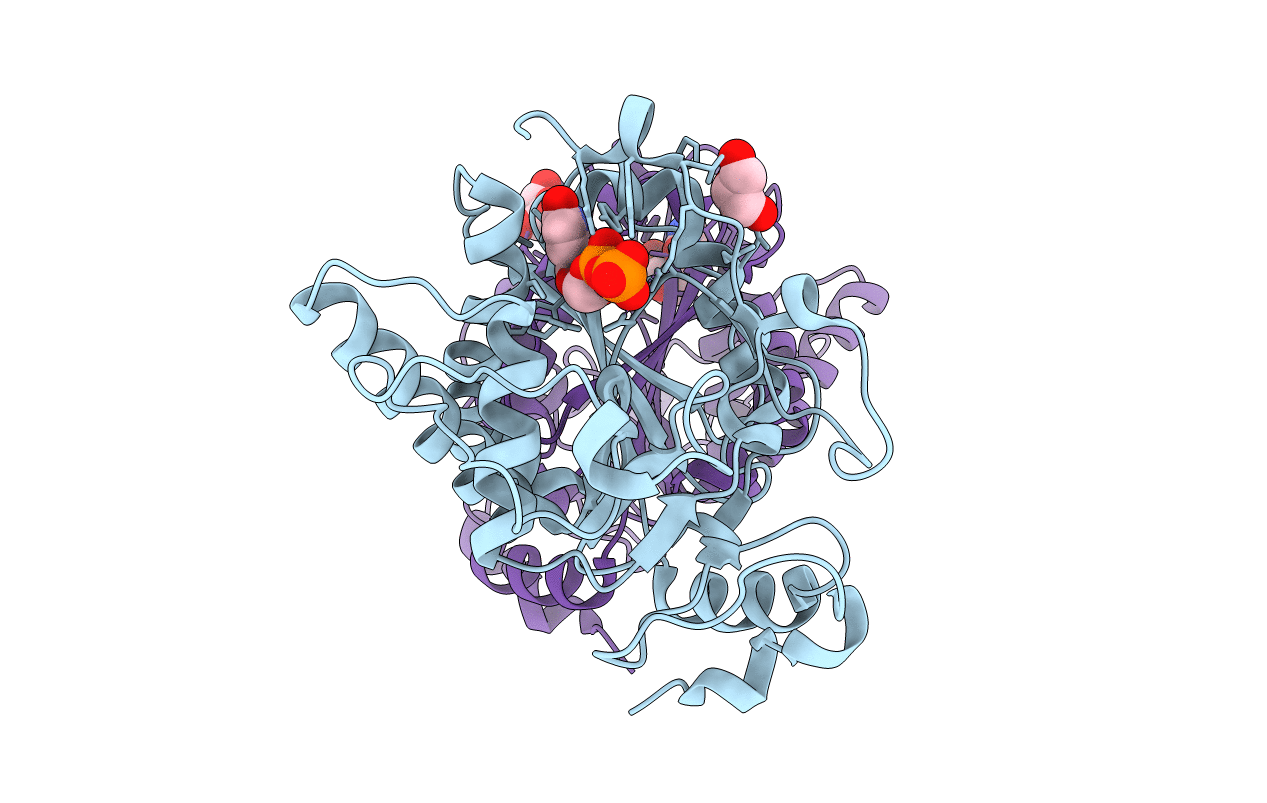
UDP-galactose:beta-galactosyl alpha-1,3-galactosyltransferase (alpha3GT) catalyzes the transfer of galactose from UDP-alpha-d-galactose into an alpha-1,3 linkage with beta-galactosyl groups in glycoconjugates. The enzyme is expressed in many mammalian species but is absent from humans, apes, and old world monkeys as a result of the mutational inactivation of the gene; in humans, a large fraction of natural antibodies are directed against its product, the alpha-galactose epitope. alpha3GT is a member of a family of metal-dependent retaining glycosyltransferases including the histo-blood group A and B synthases. A crystal structure of the catalytic domain of alpha3GT was recently reported (Gastinel, L. N., Bignon, C., Misra, A. K., Hindsgaul, O., Shaper, J. H., and Joziasse, D. H. (2001) EMBO J. 20, 638-649). However, because of the limited resolution (2.3 A) and high mobility of the atoms (as indicated by high B-factors) this structure (form I) does not provide a clear depiction of the catalytic site of the enzyme. Here we report a new, highly ordered structure for the catalytic domain of alpha3GT at 1.53-A resolution (form II). This provides a more accurate picture of the details of the catalytic site that includes a bound UDP molecule and a Mn(2+) cofactor. Significantly, in the new structure, the C-terminal segment (residues 358-368) adopts a very different, highly structured conformation and appears to form part of the active site. The properties of an Arg-365 to Lys mutant indicate that this region is important for catalysis, possibly reflecting its role in a donor substrate-induced conformational change.