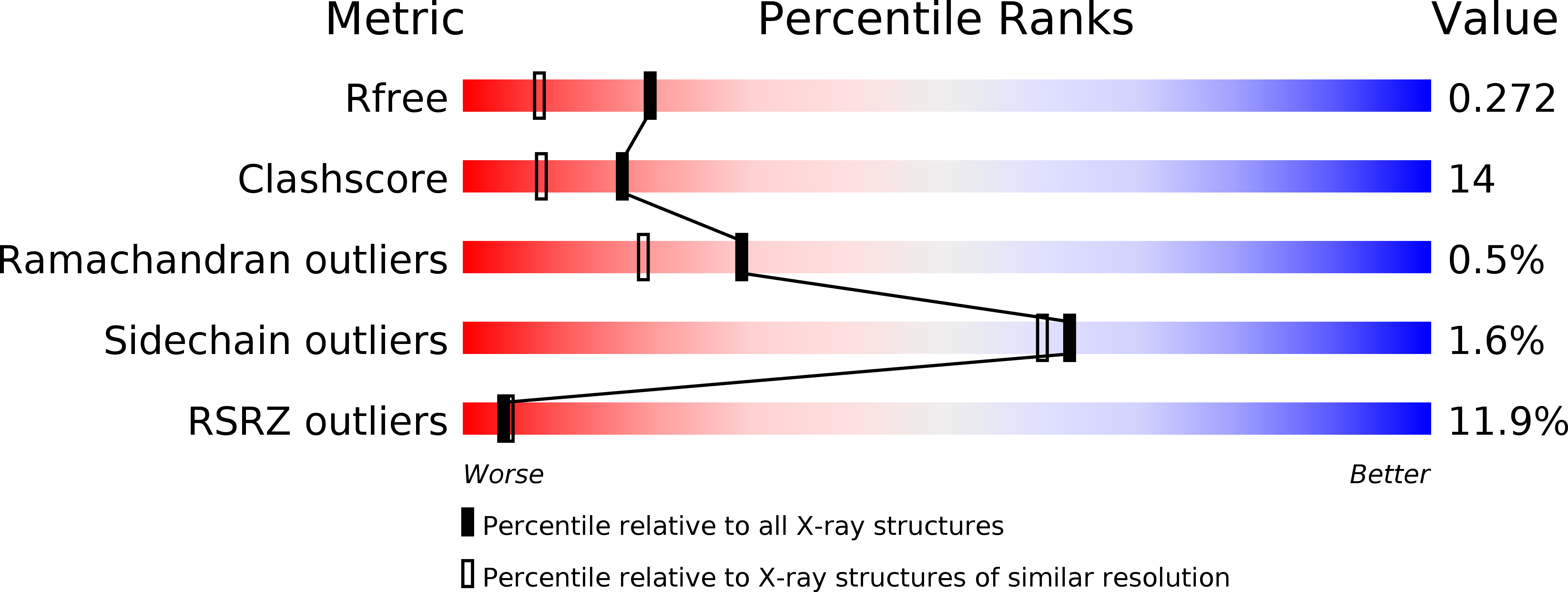
1K3S image
Deposition Date 2001-10-03
Release Date 2001-11-28
Last Version Date 2024-11-13
Entry Detail
PDB ID:
1K3S
Keywords:
Title:
Type III Secretion Chaperone SigE
Biological Source:
Source Organism:
Host Organism:
Method Details:
Experimental Method:
Resolution:
1.90 Å
R-Value Free:
0.27
R-Value Work:
0.22
R-Value Observed:
0.22
Space Group:
P 21 21 21
Macromolecular Entities
Structures with similar UniProt ID
Protein Blast
Polymer Type:polypeptide(L)
Molecule:SigE
Mutagens:none
Chain IDs:A, B
Chain Length:113
Number of Molecules:2
Biological Source:Salmonella enterica
Modified Residue
Compound ID Chain ID Parent Comp ID Details 2D Image
MSE A MET SELENOMETHIONINE
Ligand Molecules
Primary Citation
Structural and biochemical characterization of the type III secretion chaperones CesT and SigE.
Nat.Struct.Biol. 8 1031 1036 (2001)
PMID: 11685226 DOI: 10.1038/nsb717

Abstact

Several Gram-negative bacterial pathogens have evolved a type III secretion system to deliver virulence effector proteins directly into eukaryotic cells, a process essential for disease. This specialized secretion process requires customized chaperones specific for particular effector proteins. The crystal structures of the enterohemorrhagic Escherichia coli O157:H7 Tir-specific chaperone CesT and the Salmonella enterica SigD-specific chaperone SigE reveal a common overall fold and formation of homodimers. Site-directed mutagenesis suggests that variable, delocalized hydrophobic surfaces observed on the chaperone homodimers are responsible for specific binding to a particular effector protein. Isothermal titration calorimetry studies of Tir-CesT and enzymatic activity profiles of SigD-SigE indicate that the effector proteins are not globally unfolded in the presence of their cognate chaperones.

Legend

Protein

Chemical

Disease

Primary Citation of related structures