
Deposition Date
2001-10-03
Release Date
2002-10-23
Last Version Date
2023-08-16
Entry Detail
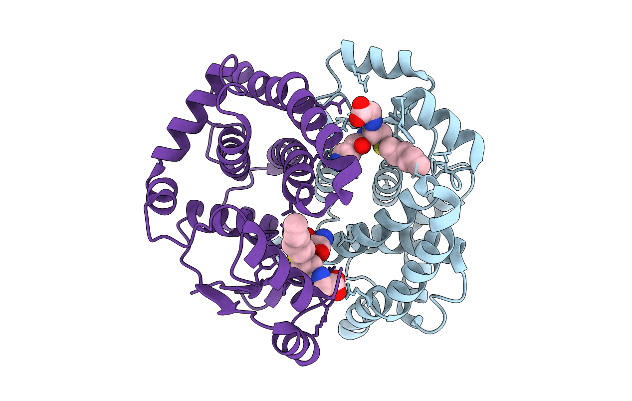
PDB ID:
1K3L
Keywords:
Title:
Crystal Structure Analysis of S-hexyl-glutathione Complex of Glutathione Transferase at 1.5 Angstroms Resolution
Biological Source:
Source Organism(s):
Homo sapiens (Taxon ID: 9606)
Expression System(s):
Method Details:
Experimental Method:
Resolution:
1.50 Å
R-Value Free:
0.24
R-Value Work:
0.15
R-Value Observed:
0.16
Space Group:
C 1 2 1
