
Deposition Date
2001-09-27
Release Date
2002-02-13
Last Version Date
2024-05-01
Entry Detail
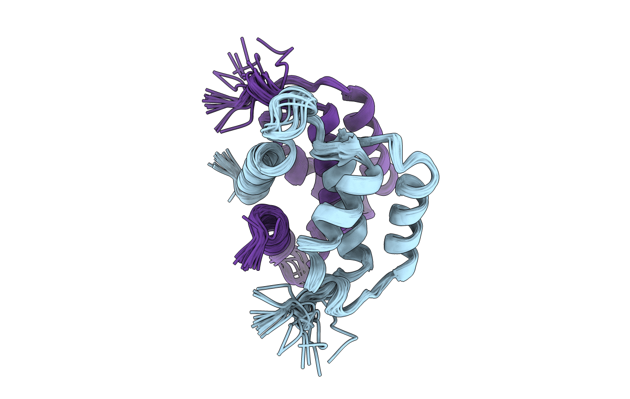
PDB ID:
1K2H
Keywords:
Title:
Three-dimensional Solution Structure of apo-S100A1.
Biological Source:
Source Organism(s):
Rattus norvegicus (Taxon ID: 10116)
Expression System(s):
Method Details:
Experimental Method:
Conformers Calculated:
75
Conformers Submitted:
20
Selection Criteria:
structures with the least restraint violations
