1K25 image
Deposition Date 2001-09-26
Release Date 2001-10-31
Last Version Date 2024-02-07
Entry Detail
PDB ID:
1K25
Title:
PBP2x from a Highly Penicillin-resistant Streptococcus pneumoniae Clinical Isolate
Biological Source:
Source Organism:
Host Organism:
Method Details:
Experimental Method:
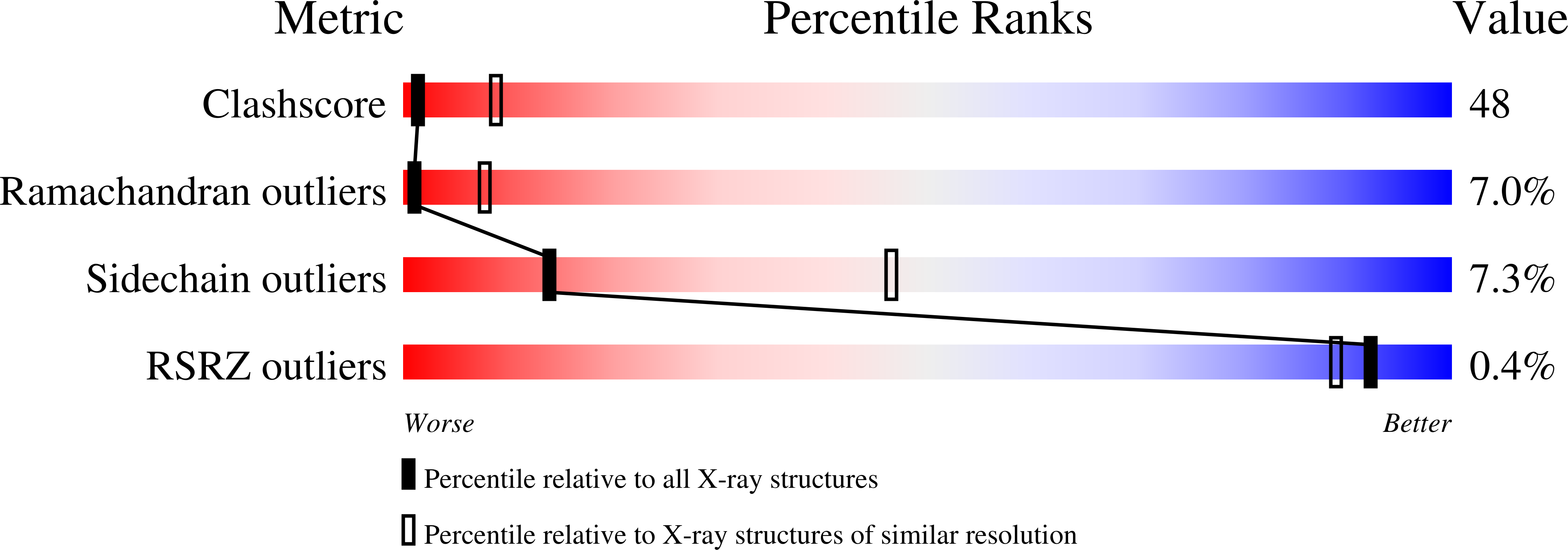
Resolution:
3.20 Å
R-Value Free:
0.31
R-Value Work:
0.23
R-Value Observed:
0.23
Space Group:
P 32
Macromolecular Entities
Structures with similar UniProt ID
Protein Blast
Polymer Type:polypeptide(L)
Molecule:low-affinity PENICILLIN-BINDING PROTEIN 2X
Gene (Uniprot):pbp2X
Mutagens:F364L
Chain IDs:A, B, C, D
Chain Length:685
Number of Molecules:4
Biological Source:Streptococcus pneumoniae
Primary Citation
Crystal structure of PBP2x from a highly penicillin-resistant Streptococcus pneumoniae clinical isolate: a mosaic framework containing 83 mutations.
J.Biol.Chem. 276 45106 45112 (2001)
PMID: 11553637 DOI: 10.1074/jbc.M107608200

Abstact

Penicillin-binding proteins (PBPs) are the main targets for beta-lactam antibiotics, such as penicillins and cephalosporins, in a wide range of bacterial species. In some Gram-positive strains, the surge of resistance to treatment with beta-lactams is primarily the result of the proliferation of mosaic PBP-encoding genes, which encode novel proteins by recombination. PBP2x is a primary resistance determinant in Streptococcus pneumoniae, and its modification is an essential step in the development of high level beta-lactam resistance. To understand such a resistance mechanism at an atomic level, we have solved the x-ray crystal structure of PBP2x from a highly penicillin-resistant clinical isolate of S. pneumoniae, Sp328, which harbors 83 mutations in the soluble region. In the proximity of the Sp328 PBP2x* active site, the Thr(338) --> Ala mutation weakens the local hydrogen bonding network, thus abrogating the stabilization of a crucial buried water molecule. In addition, the Ser(389) --> Leu and Asn(514) --> His mutations produce a destabilizing effect that generates an "open" active site. It has been suggested that peptidoglycan substrates for beta-lactam-resistant PBPs contain a large amount of abnormal, branched peptides, whereas sensitive strains tend to catalyze cross-linking of linear forms. Thus, in vivo, an "open" active site could facilitate the recognition of distinct, branched physiological substrates.

Legend

Protein

Chemical

Disease

Primary Citation of related structures