Deposition Date
2001-09-26
Release Date
2003-06-17
Last Version Date
2024-03-13
Entry Detail
PDB ID:
1K1X
Keywords:
Title:
Crystal structure of 4-alpha-glucanotransferase from thermococcus litoralis
Biological Source:
Source Organism(s):
Thermococcus litoralis (Taxon ID: 2265)
Expression System(s):
Method Details:
Experimental Method:
Resolution:
2.40 Å
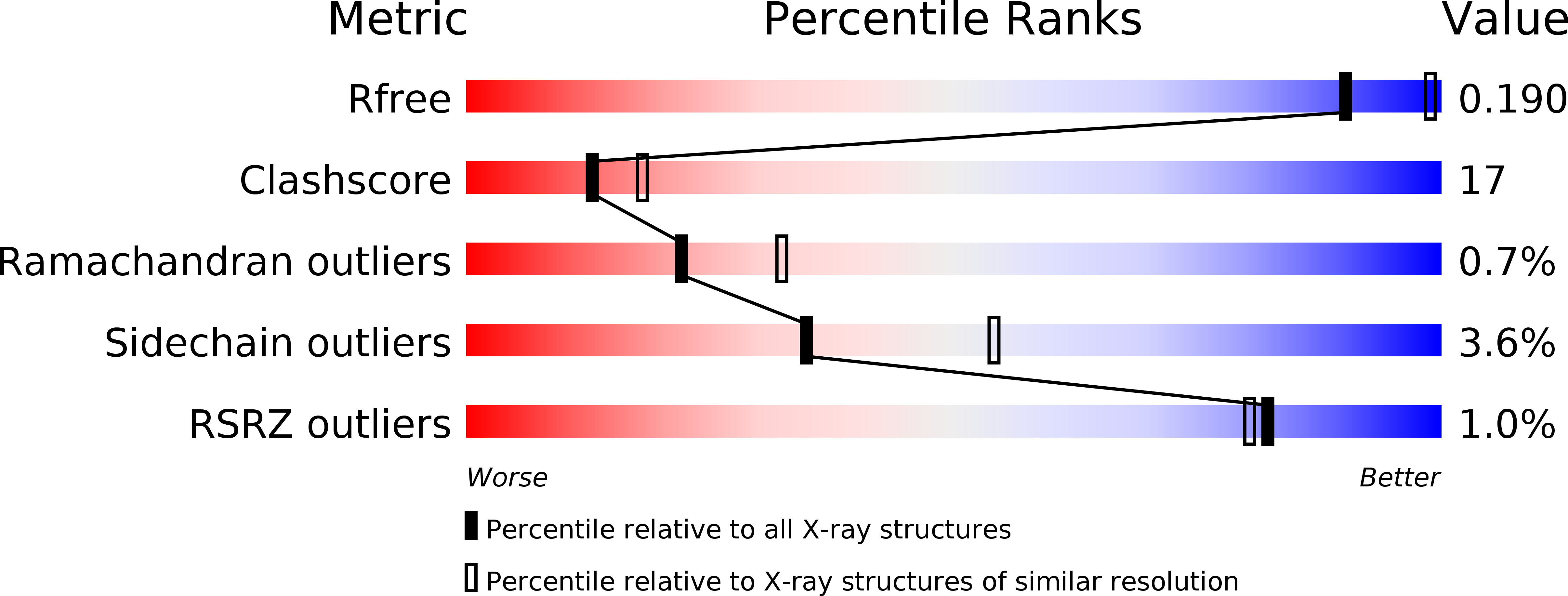
R-Value Free:
0.23
R-Value Work:
0.19
R-Value Observed:
0.19
Space Group:
P 21 21 2