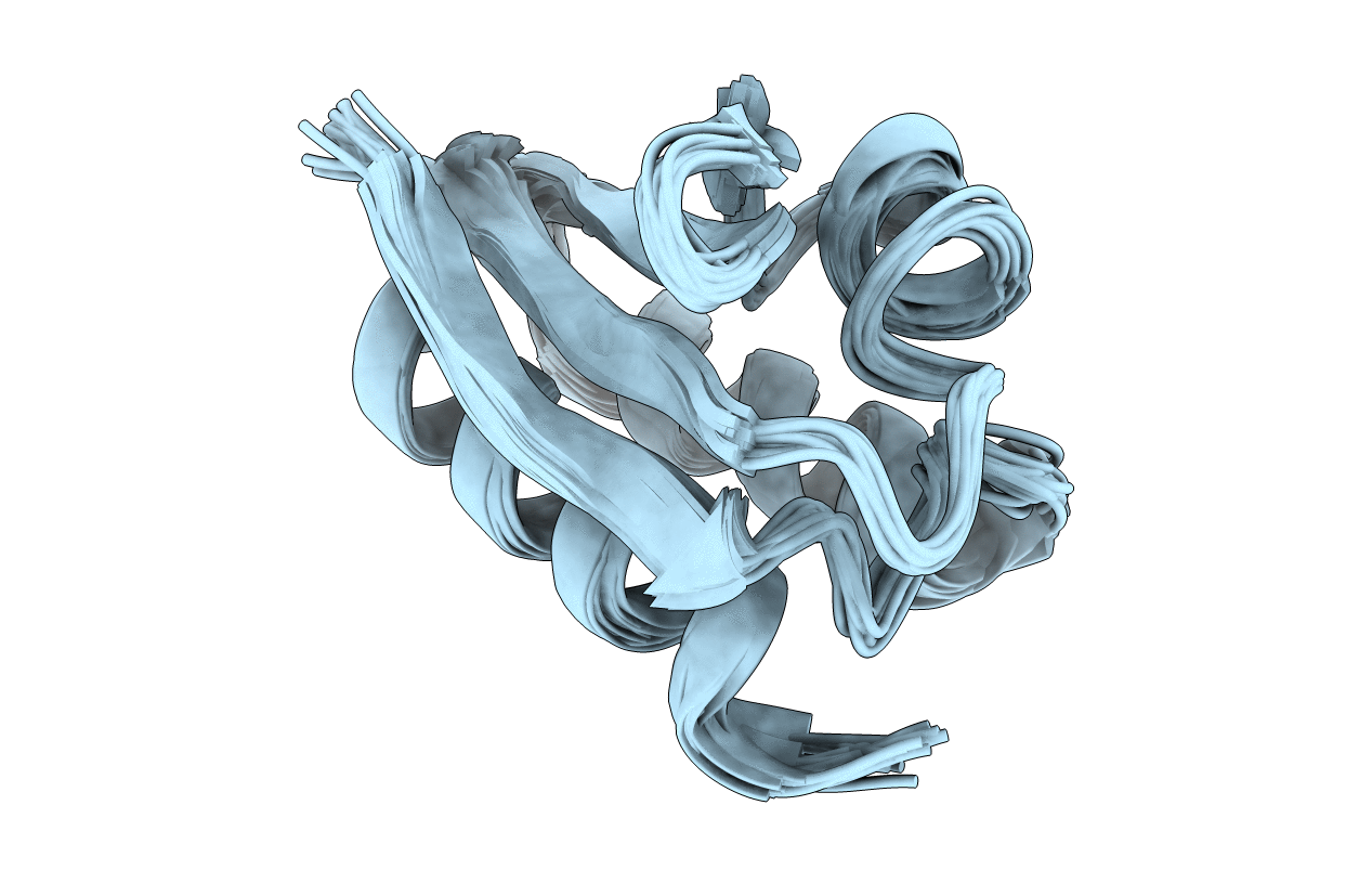
1K1C image
Deposition Date 2001-09-25
Release Date 2001-10-17
Last Version Date 2024-05-22
Entry Detail
PDB ID:
1K1C
Title:
Solution Structure of Crh, the Bacillus subtilis Catabolite Repression HPr
Biological Source:
Source Organism:
Host Organism:
Method Details:
Experimental Method:
Conformers Calculated:
250
Conformers Submitted:
24
Selection Criteria:
structures with the least restraint violations
Macromolecular Entities
Structures with similar UniProt ID
Protein Blast
Polymer Type:polypeptide(L)
Molecule:catabolite repression HPr-like protein
Gene (Uniprot):crh
Chain IDs:A
Chain Length:84
Number of Molecules:1
Biological Source:Bacillus subtilis
Ligand Molecules
Primary Citation
Solution structure and dynamics of Crh, the Bacillus subtilis catabolite repression HPr.
J.Mol.Biol. 317 131 144 (2002)
PMID: 11916384 DOI: 10.1006/jmbi.2002.5397

Abstact

The solution structure and dynamics of the Bacillus subtilis HPr-like protein, Crh, have been investigated using NMR spectroscopy. Crh exhibits high sequence identity (45 %) to the histidine-containing protein (HPr), a phospho-carrier protein of the phosphoenolpyruvate (PEP):carbohydrate phosphotransferase system, but contains no catalytic His15, the site of PEP-dependent phosphorylation in HPr. Crh also forms a mixture of monomers and dimers in solution whereas HPr is known to be monomeric. Complete backbone and side-chain assignments were obtained for the monomeric form, and 60 % of the dimer backbone resonances; allowing the identification of the Crh dimer interface from chemical-shift mapping. The conformation of Crh was determined to a precision of 0.46(+/-0.06) A for the backbone atoms, and 1.01(+/-0.08) A for the heavy atoms. The monomer structure is similar to that of known HPr 2.67(+/-0.22) A (C(alpha) rmsd), but has a few notable differences, including a change in the orientation of one of the helices (B), and a two-residue shift in beta-sheet pairing of the N-terminal strand with the beta4 strand. This shift results in a shortening of the surface loop present in HPr and consequently provides a flatter surface in the region of dimerisation contact, which may be related to the different oligomeric nature of these two proteins. A binding site of phospho-serine(P-Ser)-Crh with catabolite control protein A (CcpA) is proposed on the basis of highly conserved surface side-chains between Crh and HPr. This binding site is consistent with the model of a dimer-dimer interaction between P-Ser-Crh and CcpA. (15)N relaxation measured in the monomeric form also identified differential local mobility in the helix B which is located in the vicinity of this site.

Legend

Protein

Chemical

Disease

Primary Citation of related structures