1JZB image
Deposition Date 2001-09-14
Release Date 2002-02-27
Last Version Date 2024-11-20
Entry Detail
PDB ID:
1JZB
Keywords:
Title:
Crystal Structure of Variant 2 Scorpion Toxin from Centruroides sculpturatus Ewing
Biological Source:
Source Organism:
Method Details:
Experimental Method:
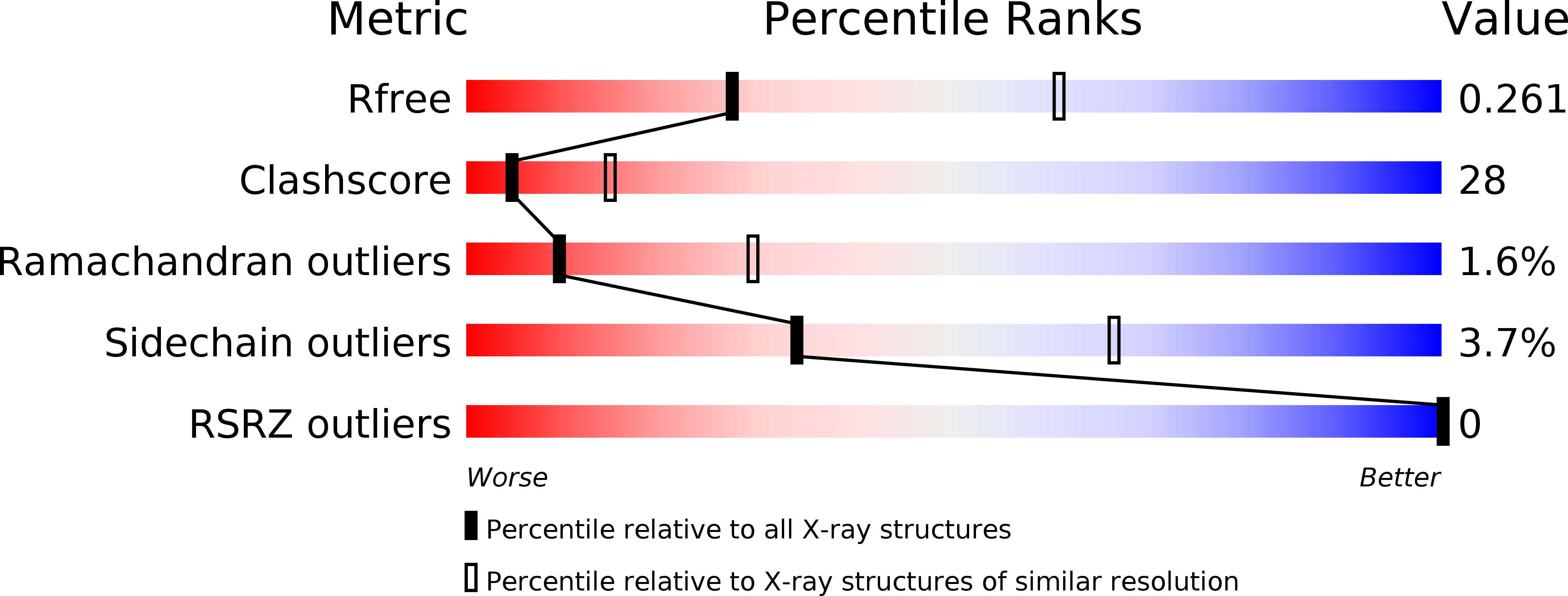
Resolution:
2.81 Å
R-Value Free:
0.27
R-Value Work:
0.23
Space Group:
P 32 2 1
Macromolecular Entities
Structures with similar UniProt ID
Protein Blast
Polymer Type:polypeptide(L)
Molecule:NEUROTOXIN 2
Chain IDs:A
Chain Length:66
Number of Molecules:1
Biological Source:Centruroides sculpturatus
Ligand Molecules
Primary Citation
Structure of variant 2 scorpion toxin from Centruroides sculpturatus Ewing.
Protein Sci. 11 479 486 (2002)
PMID: 11847271 DOI: 10.1110/ps.39202

Abstact

Centruroides sculpturatus Ewing variant 2 toxin (CsE-v2) is a neurotoxin isolated from the venom of a scorpion native to the Arizona desert. The structure of CsE-v2 was solved in two different crystal forms using a combination of molecular replacement and multiple isomorphous replacement techniques. Crystals of CsE-v2 display a temperature-dependent, reversible-phase transition near room temperature. At lower temperature the space group changes from P3(2)21 to P3(1)21 with an approximate doubling of the C-axis. The small-cell structure, which has one molecule per asymmetric unit, has an R factor of 0.229 at 2.8 A resolution. The large-cell structure has two molecules per asymmetric unit and was refined at 2.2 A resolution to an R factor of 0.255. CsE-v2 is a rigid, compact structure with four intrachain disulfide bonds. The structure is similar to other long-chain beta neurotoxins, and the largest differences occur in the last six residues. The high-resolution structure of CsE-v2 corrects an error in the reported C-terminal sequence; the terminal tripeptide sequence is Ser 64-Cys 65-Ser 66 rather than Ser 64-Ser 65-Cys 66. Comparison of CsE-v2 with long-chain alpha toxins reveals four insertions and one deletion, as well as additional residues at the N and C termini. Structural alignment of alpha and beta toxins suggests that the primary distinguishing feature between the two classes is the length of the loop between the second and third strands in a three-strand beta sheet. The shorter loop in alpha toxins exposes a critical lysine side chain, whereas the longer loop in beta toxins buries the corresponding basic residue (either arginine or lysine).

Legend

Protein

Chemical

Disease

Primary Citation of related structures
Feedback Form
Name
Email
Institute
Feedback