Deposition Date
2001-09-13
Release Date
2002-03-13
Last Version Date
2024-10-16
Entry Detail
PDB ID:
1JYR
Keywords:
Title:
Xray Structure of Grb2 SH2 Domain Complexed with a Phosphorylated Peptide
Biological Source:
Source Organism(s):
Homo sapiens (Taxon ID: 9606)
synthetic construct (Taxon ID: 32630)
synthetic construct (Taxon ID: 32630)
Expression System(s):
Method Details:
Experimental Method:
Resolution:
1.55 Å
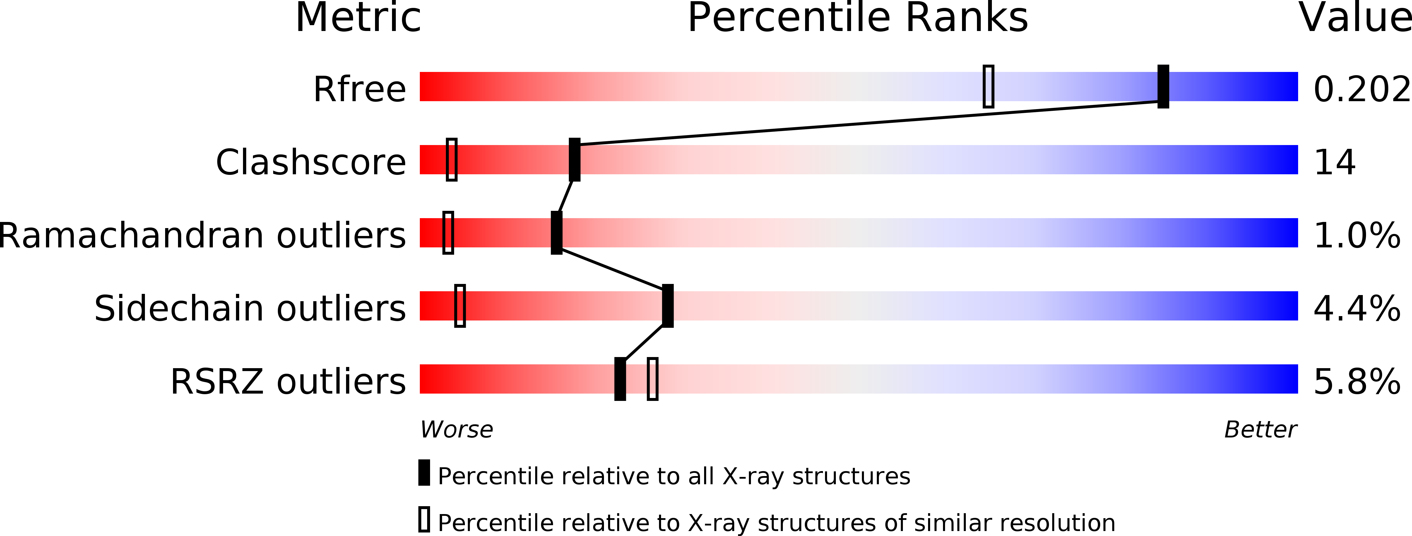
R-Value Free:
0.22
R-Value Work:
0.19
Space Group:
P 41 21 2