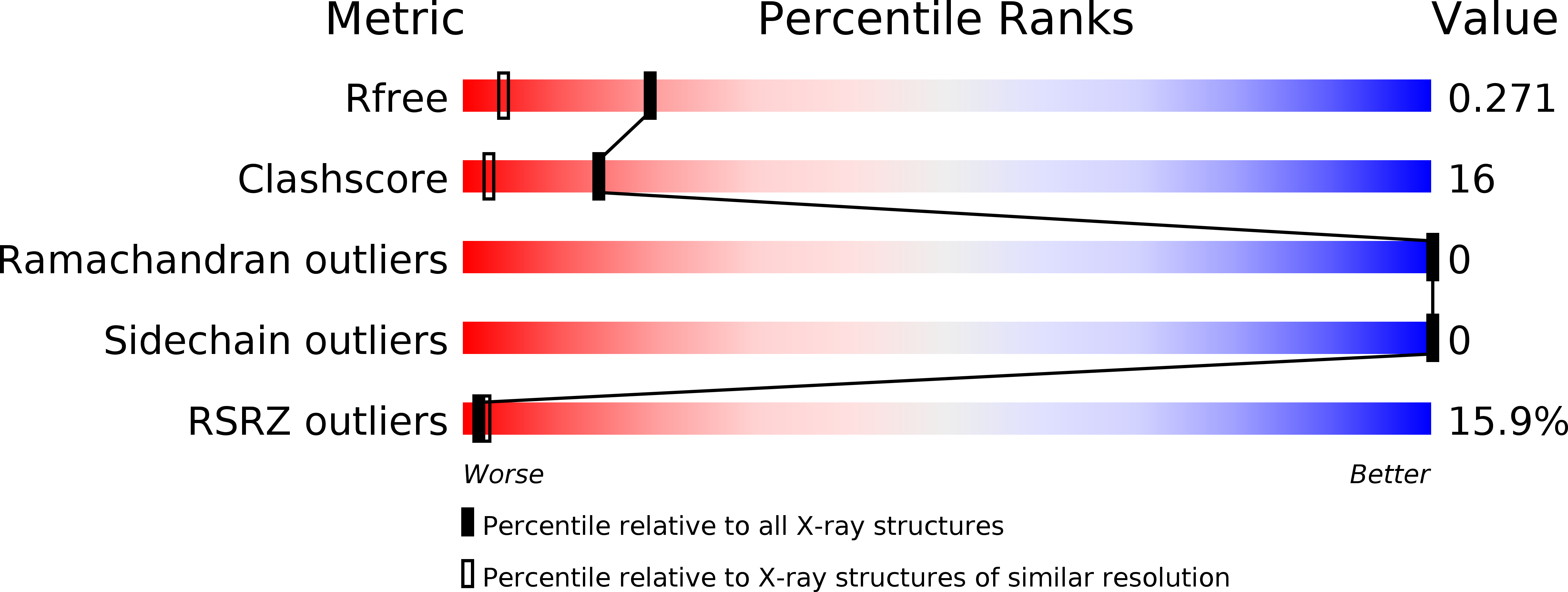
1JYA image
Deposition Date 2001-09-11
Release Date 2001-10-31
Last Version Date 2024-10-30
Entry Detail
PDB ID:
1JYA
Keywords:
Title:
Crystal Structure of SycE
Biological Source:
Source Organism:
Method Details:
Experimental Method:
Resolution:
1.74 Å
R-Value Free:
0.26
R-Value Work:
0.23
Space Group:
C 2 2 21
Macromolecular Entities
Structures with similar UniProt ID
Protein Blast
Polymer Type:polypeptide(L)
Molecule:YOPE regulator
Chain IDs:A, B
Chain Length:130
Number of Molecules:2
Biological Source:Yersinia pseudotuberculosis
Modified Residue
Compound ID Chain ID Parent Comp ID Details 2D Image
MSE A MET SELENOMETHIONINE
Primary Citation
Structure of the Yersinia type III secretory system chaperone SycE.
Nat.Struct.Biol. 8 974 978 (2001)
PMID: 11685245 DOI: 10.1038/nsb1101-974

Abstact

In the type III secretory system of bacterial pathogens, a large number of sequence-divergent but characteristically small (approximately 14-19 kDa), acidic (pI approximately 4-5) chaperone proteins have been identified. We present the 1.74 A resolution crystal structure of the Yersinia pseudotuberculosis chaperone SycE, whose action in promoting translocation of YopE into host macrophages is essential to Yersinia pathogenesis. SycE, a compact, globular dimer with a novel fold, has two large hydrophobic surface patches that may form binding sites for YopE or other type III components. These patches are formed by structurally key residues that are conserved among many chaperones, suggesting shared structural and functional relationships. A negative electrostatic potential covers almost the entire surface of SycE and is likely conserved in character, but not in detail, among chaperones. The structure provides the first structural insights into possible modes of action of SycE and type III chaperones in general.

Legend

Protein

Chemical

Disease

Primary Citation of related structures