1JY2 image
Deposition Date 2001-09-10
Release Date 2001-10-17
Last Version Date 2024-11-06
Entry Detail
PDB ID:
1JY2
Keywords:
Title:
Crystal Structure of the Central Region of Bovine Fibrinogen (E5 fragment) at 1.4 Angstroms Resolution
Biological Source:
Source Organism:
Bos taurus (Taxon ID: 9913)
Method Details:
Experimental Method:
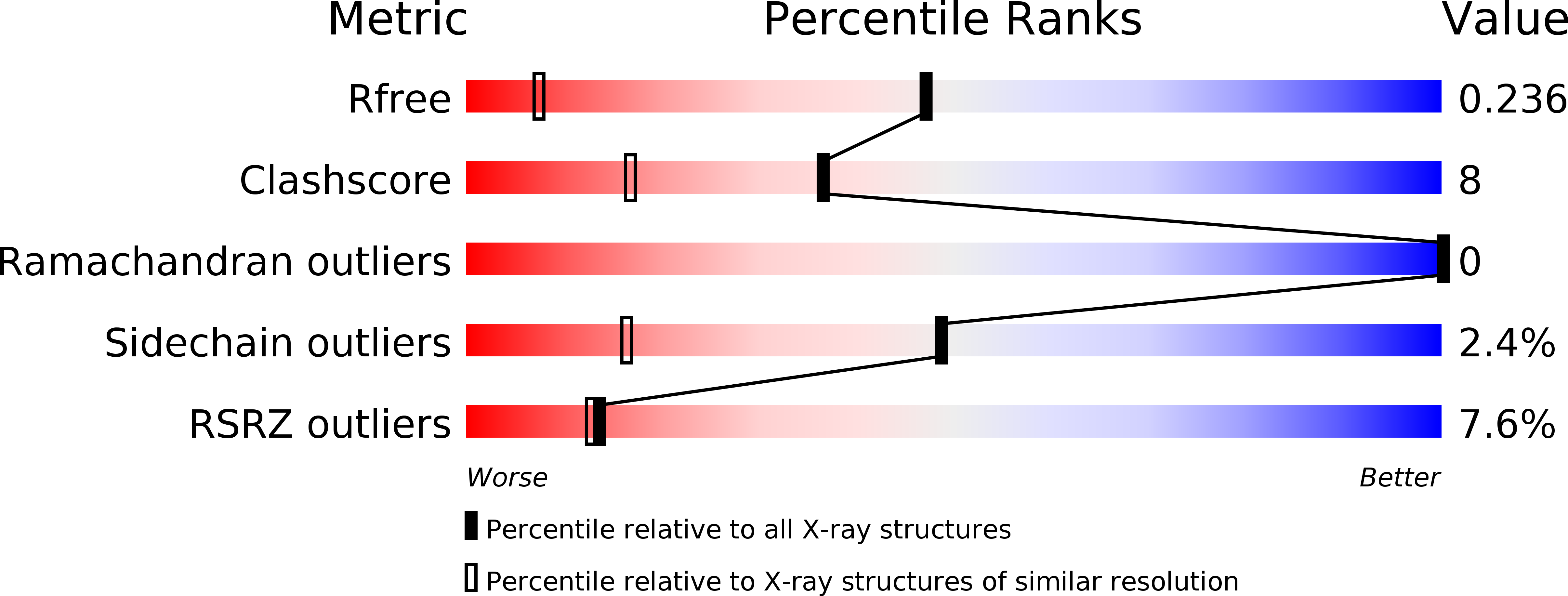
Resolution:
1.40 Å
R-Value Free:
0.23
R-Value Work:
0.21
Space Group:
P 1 21 1
Macromolecular Entities
Polymer Type:polypeptide(L)
Molecule:FIBRINOGEN ALPHA CHAIN
Chain IDs:A (auth: N), D (auth: Q)
Chain Length:53
Number of Molecules:2
Biological Source:Bos taurus
Polymer Type:polypeptide(L)
Molecule:FIBRINOGEN BETA CHAIN
Gene (Uniprot):FGB
Chain IDs:B (auth: O), E (auth: R)
Chain Length:56
Number of Molecules:2
Biological Source:Bos taurus
Polymer Type:polypeptide(L)
Molecule:FIBRINOGEN GAMMA-B CHAIN
Gene (Uniprot):FGG
Chain IDs:C (auth: P), F (auth: S)
Chain Length:48
Number of Molecules:2
Biological Source:Bos taurus
Primary Citation
Crystal structure of the central region of bovine fibrinogen (E5 fragment) at 1.4-A resolution.
Proc.Natl.Acad.Sci.USA 98 11967 11972 (2001)
PMID: 11593005 DOI: 10.1073/pnas.211439798

Abstact

The high-resolution crystal structure of the N-terminal central region of bovine fibrinogen (a 35-kDa E(5) fragment) reveals a remarkable dimeric design. The two halves of the molecule bond together at the center in an extensive molecular "handshake" by using both disulfide linkages and noncovalent contacts. On one face of the fragment, the Aalpha and Bbeta chains from the two monomers form a funnel-shaped domain with an unusual hydrophobic cavity; here, on each of the two outer sides there appears to be a binding site for thrombin. On the opposite face, the N-terminal gamma chains fold into a separate domain. Despite the chemical identity of the two halves of fibrinogen, an unusual pair of adjacent disulfide bonds locally constrain the two gamma chains to adopt different conformations. The striking asymmetry of this domain may promote the known supercoiling of the protofibrils in fibrin. This information on the detailed topology of the E(5) fragment permits the construction of a more detailed model than previously possible for the critical trimolecular junction of the protofibril in fibrin.

Legend

Protein

Chemical

Disease

Primary Citation of related structures