
Deposition Date
2001-09-05
Release Date
2001-11-07
Last Version Date
2024-10-30
Entry Detail
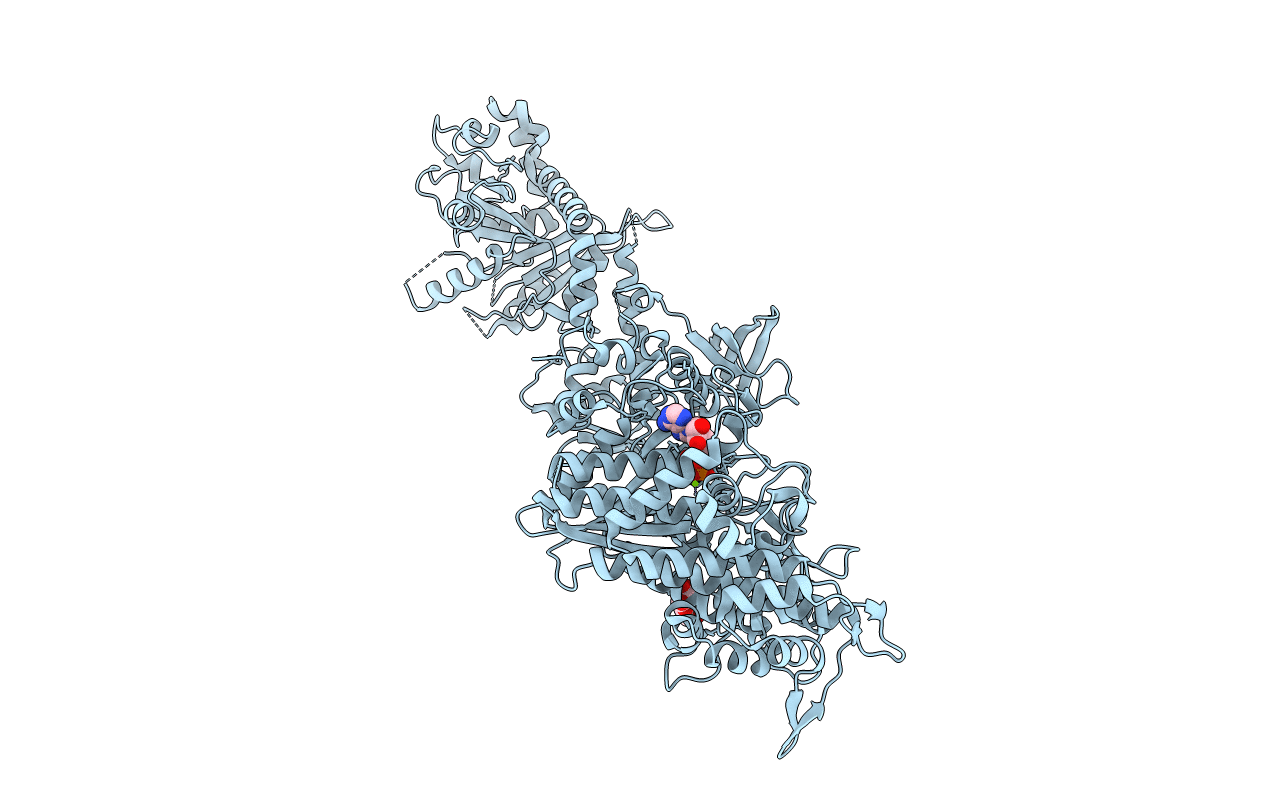
PDB ID:
1JX2
Keywords:
Title:
CRYSTAL STRUCTURE OF THE NUCLEOTIDE-FREE DYNAMIN A GTPASE DOMAIN, DETERMINED AS MYOSIN FUSION
Biological Source:
Source Organism(s):
Dictyostelium discoideum (Taxon ID: 44689)
Expression System(s):
Method Details:
Experimental Method:
Resolution:
2.30 Å
R-Value Free:
0.25
R-Value Work:
0.19
R-Value Observed:
0.19
Space Group:
P 1 21 1
