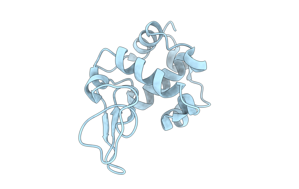
1JWR image
Deposition Date 2001-09-05
Release Date 2001-09-19
Last Version Date 2024-10-23
Entry Detail
PDB ID:
1JWR
Keywords:
Title:
Crystal structure of human lysozyme at 100 K
Biological Source:
Source Organism:
Homo sapiens (Taxon ID: 9606)
Host Organism:
Method Details:
Experimental Method:
Resolution:
1.40 Å
R-Value Free:
0.21
R-Value Work:
0.17
Space Group:
P 21 21 21
Macromolecular Entities
Structures with similar UniProt ID
Protein Blast
Polymer Type:polypeptide(L)
Molecule:lysozyme
Gene (Uniprot):LYZ
Chain IDs:A
Chain Length:130
Number of Molecules:1
Biological Source:Homo sapiens
Primary Citation
Hydration structure of human lysozyme investigated by molecular dynamics simulation and cryogenic X-ray crystal structure analyses: on the correlation between crystal water sites, solvent density, and solvent dipole
J.Comput.Chem. 23 1323 1336 (2002)
PMID: 12214315 DOI: 10.1002/jcc.10100

Abstact

The hydration structure of human lysozyme was studied with cryogenic X-ray diffraction experiment and molecular dynamics simulations. The crystal structure analysis at a resolution of 1.4 A provided 405 crystal water molecules around the enzyme. In the simulations at 300 K, the crystal structure was immersed in explicit water molecules. We examined correlations between crystal water sites and two physical quantities calculated from the 1-ns simulation trajectories: the solvent density reflecting the time-averaged distribution of water molecules, and the solvent dipole measuring the orientational ordering of water molecules around the enzyme. The local high solvent density sites were consistent with the crystal water sites, and better correlation was observed around surface residues with smaller conformational fluctuations during the simulations. Solvent dipoles around those sites exhibited coherent and persistent ordering, indicating that the hydration water molecules at the crystal water sites were highly oriented through the interactions with hydrophilic residues. Those water molecules restrained the orientational motions of adjoining water molecules and induced a solvent dipole field, which was persistent during the simulations around the enzyme. The coherent ordering was particularly prominent in and around the active site cleft of the enzyme. Because the ordering was significant up to the third to fourth solvent layer region from the enzyme surface, the coherently ordered solvent dipoles likely contributed to the molecular recognition of the enzyme in a long-distance range. The present work may provide a new approach combining computational and the experimental studies to understand protein hydration.

Legend

Protein

Chemical

Disease

Primary Citation of related structures