1JWD image
Deposition Date 2001-09-04
Release Date 2002-03-27
Last Version Date 2024-05-22
Entry Detail
PDB ID:
1JWD
Title:
Ca2+-induced Structural Changes in Calcyclin: High-resolution Solution Structure of Ca2+-bound Calcyclin.
Biological Source:
Source Organism:
Host Organism:
Method Details:
Experimental Method:
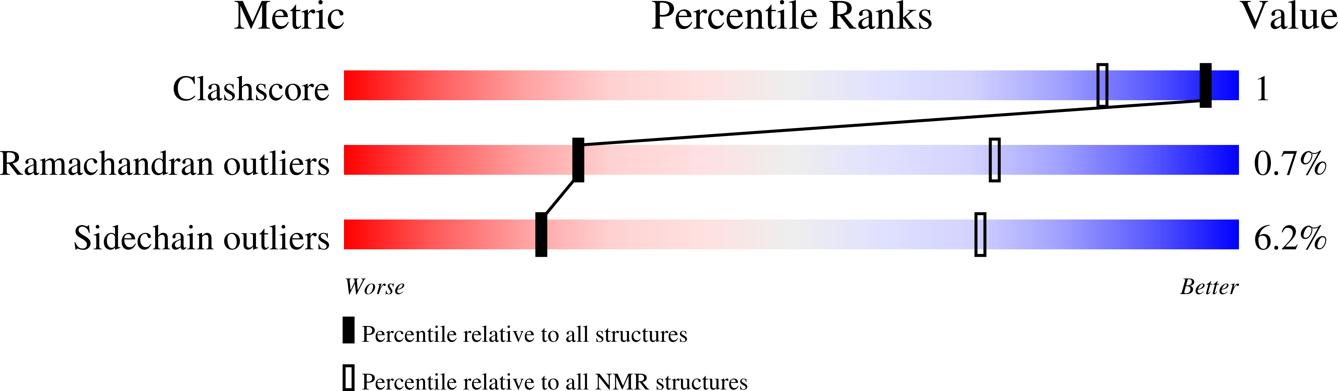
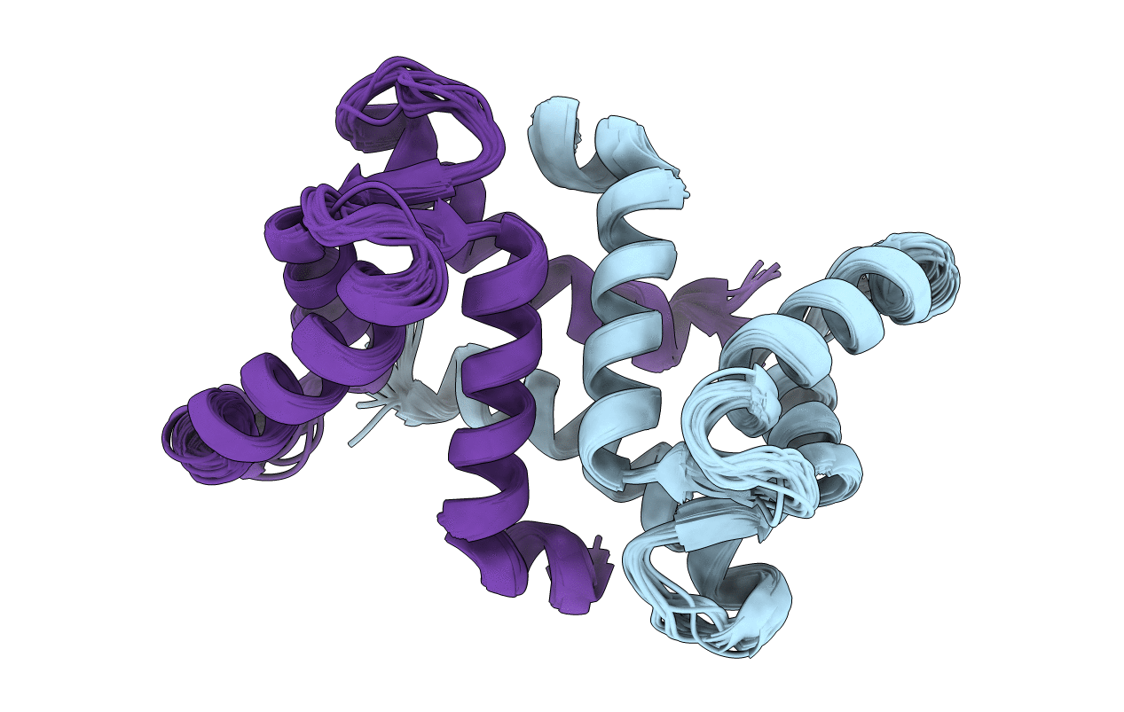
Conformers Calculated:
100
Conformers Submitted:
22
Selection Criteria:
The program Findfam was used to establish that the number of structures required to accurately represent the ensemble was less than 22 (the number selected to represent previous S100A6 ensembles). Structures were ordered by lowest restraint violations, then accepted if total molecular energy and each contributing term was within two standard deviations of the mean. The 22 structures with least restraint violations (energy penalty and magnitude of largest violation) all met these criteria.
Macromolecular Entities
Structures with similar UniProt ID
Protein Blast
Polymer Type:polypeptide(L)
Molecule:Calcyclin
Gene (Uniprot):S100A6
Chain IDs:A, B
Chain Length:90
Number of Molecules:2
Biological Source:Oryctolagus cuniculus
Ligand Molecules
Primary Citation
A structural basis for S100 protein specificity derived from comparative analysis of apo and Ca(2+)-calcyclin
J.Mol.Biol. 317 279 290 (2002)
PMID: 11902843 DOI: 10.1006/jmbi.2002.5421

Abstact

Calcyclin is a homodimeric protein belonging to the S100 subfamily of EF-hand Ca(2+)-binding proteins, which function in Ca(2+) signal transduction processes. A refined high-resolution solution structure of Ca(2+)-bound rabbit calcyclin has been determined by heteronuclear solution NMR. In order to understand the Ca(2+)-induced structural changes in S100 proteins, in-depth comparative structural analyses were used to compare the apo and Ca(2+)-bound states of calcyclin, the closely related S100B, and the prototypical Ca(2+)-sensor protein calmodulin. Upon Ca(2+) binding, the position and orientation of helix III in the second EF-hand is altered, whereas the rest of the protein, including the dimer interface, remains virtually unchanged. This Ca(2+)-induced structural change is much less drastic than the "opening" of the globular EF-hand domains that occurs in classical Ca(2+) sensors, such as calmodulin. Using homology models of calcyclin based on S100B, a binding site in calcyclin has been proposed for the N-terminal domain of annexin XI and the C-terminal domain of the neuronal calcyclin-binding protein. The structural basis for the specificity of S100 proteins is discussed in terms of the variation in sequence of critical contact residues in the common S100 target-binding site.

Legend

Protein

Chemical

Disease

Primary Citation of related structures