1JW6 image
Deposition Date 2001-09-02
Release Date 2001-09-26
Last Version Date 2023-10-25
Entry Detail
PDB ID:
1JW6
Title:
Crystal Structure of the Complex of Concanavalin A and Hexapeptide
Biological Source:
Source Organism:
Method Details:
Experimental Method:
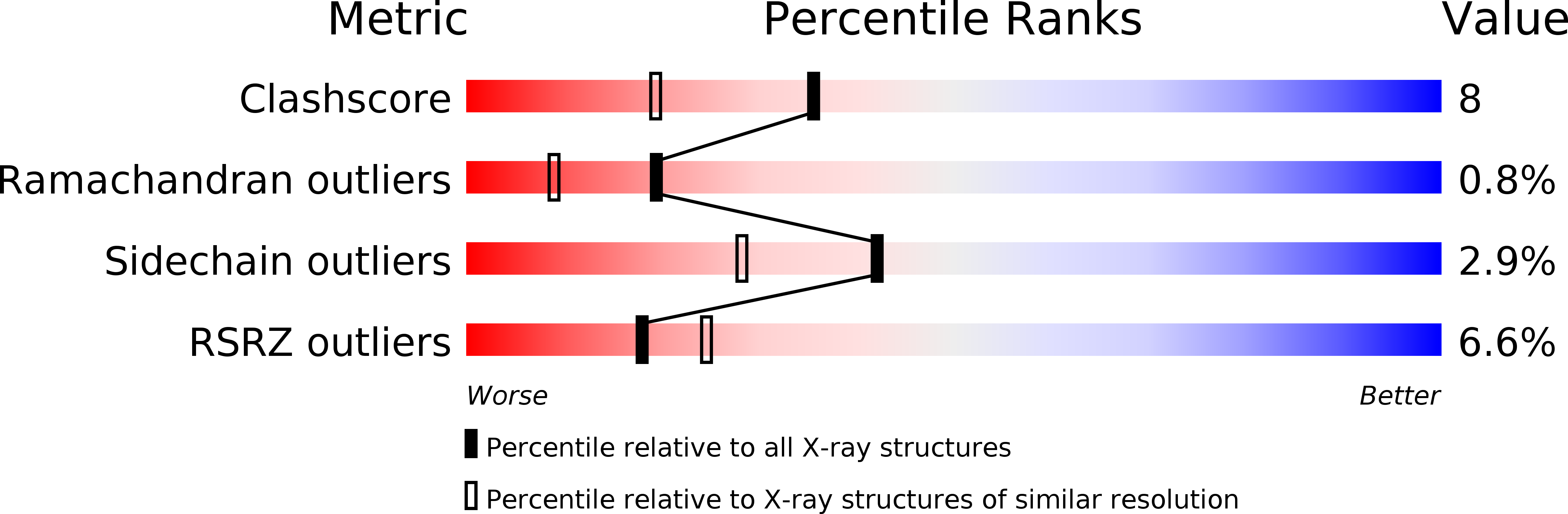
Resolution:
1.93 Å
R-Value Free:
0.25
R-Value Work:
0.19
R-Value Observed:
0.21
Space Group:
I 2 2 2
Macromolecular Entities
Polymer Type:polypeptide(L)
Molecule:Concanavalin A
Chain IDs:A
Chain Length:237
Number of Molecules:1
Biological Source:Canavalia ensiformis
Polymer Type:polypeptide(L)
Molecule:PROTEIN (6-MER)
Chain IDs:B
Chain Length:6
Number of Molecules:1
Biological Source:synthetic construct
Primary Citation
Crystal structure of the complex of concanavalin A and hexapeptide.
J.Protein Chem. 20 423 429 (2001)
PMID: 11732694 DOI: 10.1023/A:1012289022412

Abstact

The X-ray structure analysis of a cross-linked crystal of concanavalin A soaked with a hexapeptide molecule as a probe molecule showed an electron density corresponding to full occupation in the binding pocket. The site lies on the surface of concanavalin A and is surrounded by three symmetry-related molecules. The crystal structure of the hexapeptide complex was refined at 1.93-A resolution, to an R-factor of 19% (Rfree factor of 25%), with an RMS deviation in bond distances of 0.01 A. The model includes all 237 residues of concanavalin A, one manganese ion, one calcium ion, 95 water molecules, one glutaraldehyde molecule, one isopropanol molecule, and one hexapeptide molecule. This X-ray structure analysis also provides an approach to mapping the binding surface of crystalline protein with a probe molecule that is dissolved in the mixture of organic solvent with water or in neat organic solvent but is hardly dissolved in aqueous solution.

Legend

Protein

Chemical

Disease

Primary Citation of related structures