1JVB image
Deposition Date 2001-08-29
Release Date 2002-08-29
Last Version Date 2024-11-13
Entry Detail
PDB ID:
1JVB
Keywords:
Title:
ALCOHOL DEHYDROGENASE FROM THE ARCHAEON SULFOLOBUS SOLFATARICUS
Biological Source:
Source Organism:
Host Organism:
Method Details:
Experimental Method:
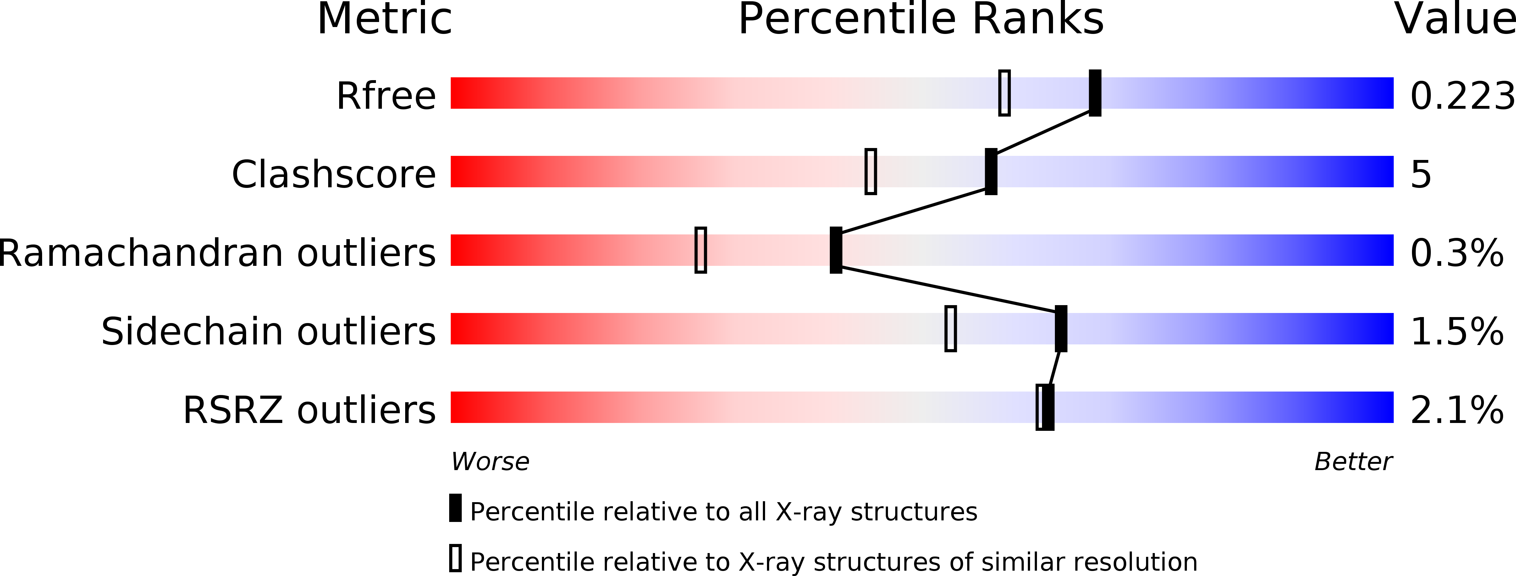
Resolution:
1.85 Å
R-Value Free:
0.22
R-Value Work:
0.19
Space Group:
I 41 2 2
Macromolecular Entities
Polymer Type:polypeptide(L)
Molecule:NAD(H)-DEPENDENT ALCOHOL DEHYDROGENASE
Gene (Uniprot):adh
Chain IDs:A
Chain Length:347
Number of Molecules:1
Biological Source:Sulfolobus solfataricus
Modified Residue
Compound ID Chain ID Parent Comp ID Details 2D Image
MSE A MET SELENOMETHIONINE
Ligand Molecules
Primary Citation
Crystal structure of the alcohol dehydrogenase from the hyperthermophilic archaeon Sulfolobus solfataricus at 1.85 A resolution.
J.Mol.Biol. 318 463 477 (2002)
PMID: 12051852 DOI: 10.1016/S0022-2836(02)00088-8

Abstact

The crystal structure of a medium-chain NAD(H)-dependent alcohol dehydrogenase (ADH) from an archaeon has been solved by multiwavelength anomalous diffraction, using a selenomethionine-substituted enzyme. The protein (SsADH), extracted from the hyperthermophilic organism Sulfolobus solfataricus, is a homo-tetramer with a crystallographic 222 symmetry. Despite the low level of sequence identity, the overall fold of the monomer is similar to that of the other homologous ADHs of known structure. However, a significant difference is the orientation of the catalytic domain relative to the coenzyme-binding domain that results in a larger interdomain cleft. At the bottom of this cleft, the catalytic zinc ion is coordinated tetrahedrally and lacks the zinc-bound water molecule that is usually found in ADH apoform structures. The fourth coordination position is indeed occupied by a Glu residue, as found in bacterial tetrameric ADHs. Other differences are found in the architecture of the substrate pocket whose entrance is more restricted than in other ADHs. SsADH is the first tetrameric ADH X-ray structure containing a second zinc ion playing a structural role. This latter metal ion shows a peculiar coordination, with a glutamic acid residue replacing one of the four cysteine ligands that are highly conserved throughout the structural zinc-containing dimeric ADHs.

Legend

Protein

Chemical

Disease

Primary Citation of related structures