1JUS image
Entry Detail
PDB ID:
1JUS
Keywords:
Title:
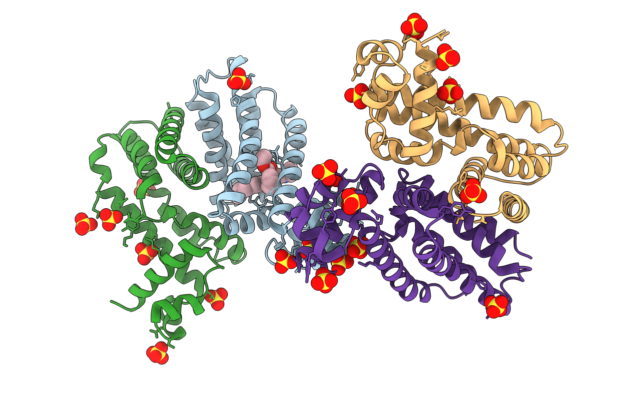
Crystal structure of the multidrug binding transcriptional repressor QacR bound to rhodamine 6G
Biological Source:
Source Organism:
Host Organism:
PDB Version:
Deposition Date:
2001-08-27
Release Date:
2001-12-12
Method Details:
Experimental Method:
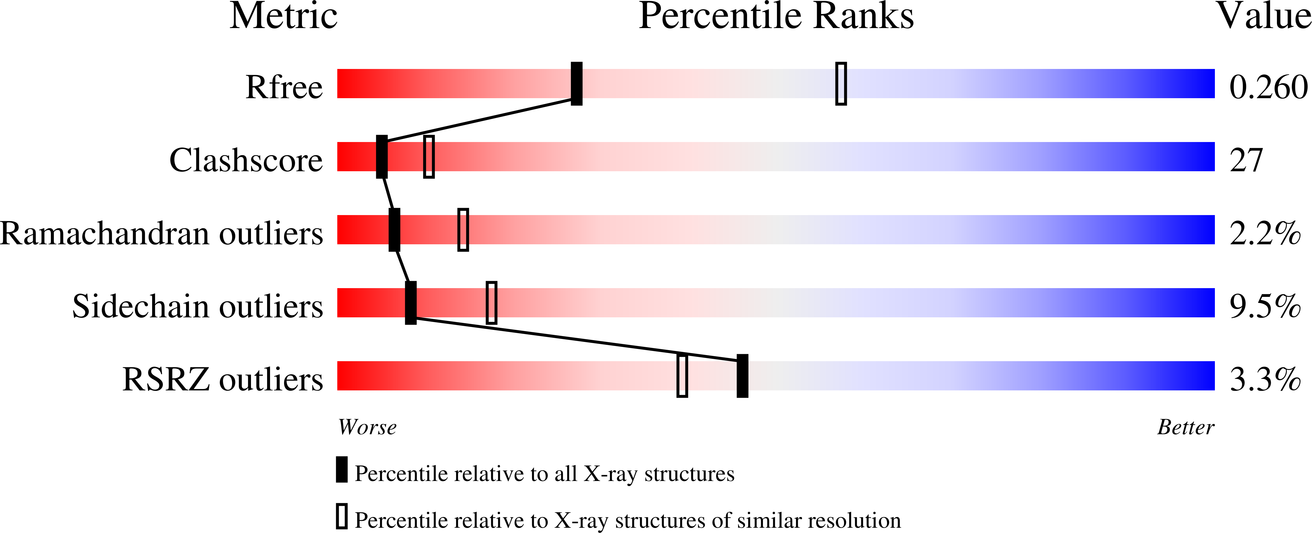
Resolution:
2.84 Å
R-Value Free:
0.27
R-Value Work:
0.23
Space Group:
P 42 21 2
Macromolecular Entities
Polymer Type:polypeptide(L)
Description:HYPOTHETICAL TRANSCRIPTIONAL REGULATOR IN QACA 5'REGION
Mutations:C72A, C141S
Chain IDs:A (auth: B), B (auth: D), C (auth: A), D (auth: E)
Chain Length:194
Number of Molecules:4
Biological Source:Staphylococcus aureus
Modified Residue
Compound ID Chain ID Parent Comp ID Details 2D Image
MSE A MET SELENOMETHIONINE
Primary Citation
Structural mechanisms of QacR induction and multidrug recognition.
Science 294 2158 2163 (2001)
PMID: 11739955 DOI: 10.1126/science.1066020

Abstact

The Staphylococcus aureus multidrug binding protein QacR represses transcription of the qacA multidrug transporter gene and is induced by structurally diverse cationic lipophilic drugs. Here, we report the crystal structures of six QacR-drug complexes. Compared to the DNA bound structure, drug binding elicits a coil-to-helix transition that causes induction and creates an expansive multidrug-binding pocket, containing four glutamates and multiple aromatic and polar residues. These structures indicate the presence of separate but linked drug-binding sites within a single protein. This multisite drug-binding mechanism is consonant with studies on multidrug resistance transporters.

Legend

Protein

Chemical

Disease

Primary Citation of related structures