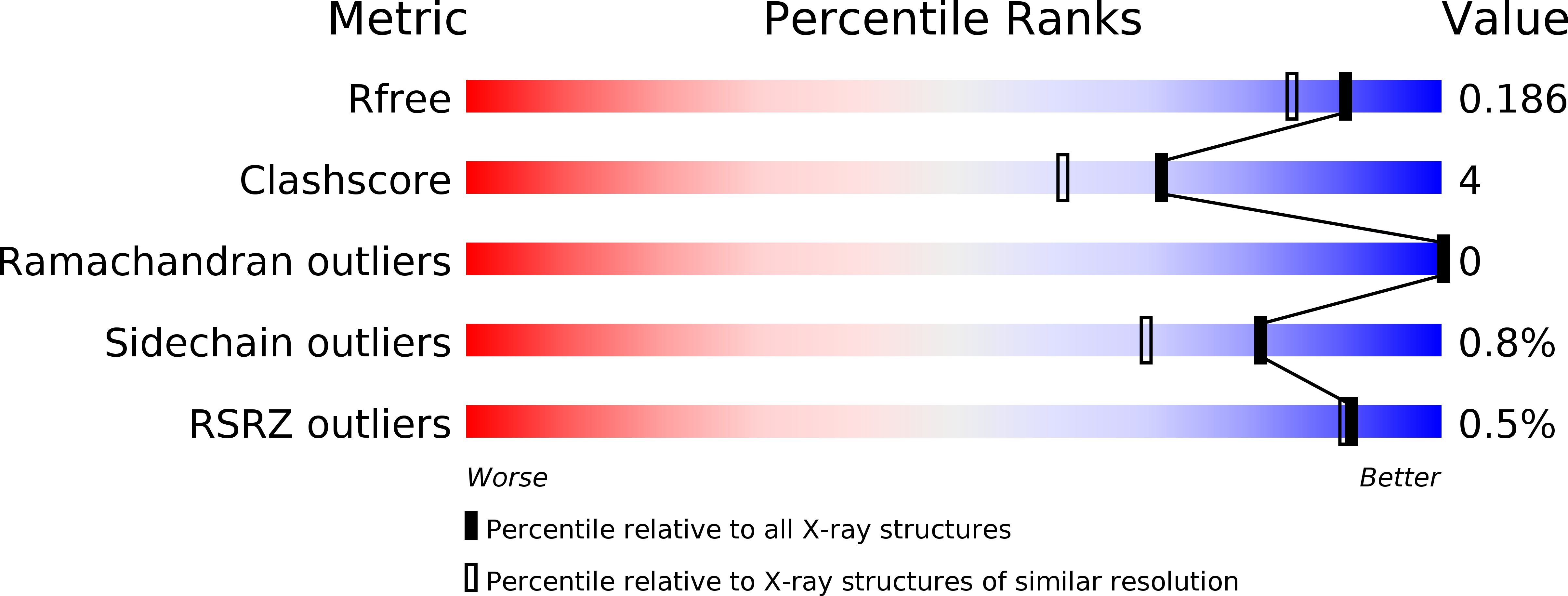
1JUH image
Entry Detail
PDB ID:
1JUH
Keywords:
Title:
Crystal Structure of Quercetin 2,3-dioxygenase
Biological Source:
Source Organism:
PDB Version:
Deposition Date:
2001-08-24
Release Date:
2002-05-22
Method Details:
Experimental Method:
Resolution:
1.60 Å
R-Value Free:
0.18
R-Value Work:
0.16
R-Value Observed:
0.16
Space Group:
P 1 21 1
Macromolecular Entities
Polymer Type:polypeptide(L)
Description:quercetin 2,3-dioxygenase
Chain IDs:A, B, C, D
Chain Length:350
Number of Molecules:4
Biological Source:Aspergillus japonicus
Modified Residue
Compound ID Chain ID Parent Comp ID Details 2D Image
ASN A ASN GLYCOSYLATION SITE
Primary Citation
Crystal structure of the copper-containing quercetin 2,3-dioxygenase from Aspergillus japonicus.
Structure 10 259 268 (2002)
PMID: 11839311 DOI: 10.1016/s0969-2126(02)00704-9

Abstact

Quercetin 2,3-dioxygenase is a copper-containing enzyme that catalyzes the insertion of molecular oxygen into polyphenolic flavonols. Dioxygenation catalyzed by iron-containing enzymes has been studied extensively, but dioxygenases employing other metal cofactors are poorly understood. We determined the crystal structure of quercetin 2,3-dioxygenase at 1.6 A resolution. The enzyme forms homodimers, which are stabilized by an N-linked heptasaccharide at the dimer interface. The mononuclear type 2 copper center displays two distinct geometries: a distorted tetrahedral coordination, formed by His66, His68, His112, and a water molecule, and a distorted trigonal bipyramidal environment, which additionally comprises Glu73. Manual docking of the substrate quercetin into the active site showed that the different geometries of the copper site might be of catalytic importance.

Legend

Protein

Chemical

Disease

Primary Citation of related structures