
Deposition Date
1996-07-03
Release Date
1997-01-11
Last Version Date
2024-11-20
Entry Detail
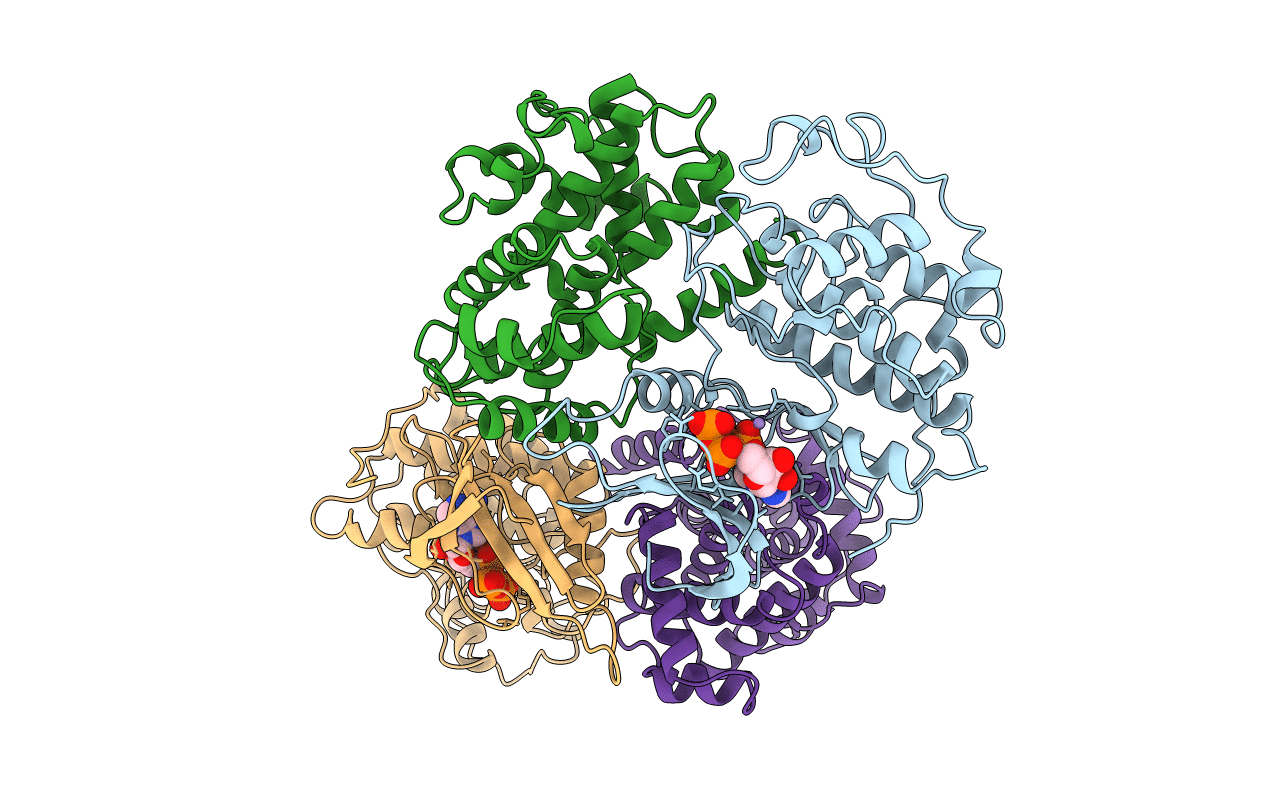
PDB ID:
1JST
Keywords:
Title:
PHOSPHORYLATED CYCLIN-DEPENDENT KINASE-2 BOUND TO CYCLIN A
Biological Source:
Source Organism(s):
Homo sapiens (Taxon ID: 9606)
Expression System(s):
Method Details:
Experimental Method:
Resolution:
2.60 Å
R-Value Work:
0.20
Space Group:
P 21 21 21
